

A Case of Multiple Evanescent White Dot Syndrome in a Myopic Male
 Save as PDF
This paper details a case of MEWDS in a seldom-affected gender and emphasizes the benefit of early, multimodal diagnostic imaging to confirm the…
Read More


 Save as PDF
This paper details a case of MEWDS in a seldom-affected gender and emphasizes the benefit of early, multimodal diagnostic imaging to confirm the…
Read More Save as PDF
Best vitelliform macular dystrophy (BVMD) is a relatively common inherited retinal disease caused by one of hundreds of known mutations in the…
Read More Save as PDF
Sclerochoroidal calcifications are uncommon, benign choroidal lesions of calcific deposition occurring at the oblique extraocular muscle insertion.
Read More Save as PDF
Macular telangiectasia (MacTel) type 2 frequently presents with intraretinal crystals along with the characteristic abnormal vessel development.
Read More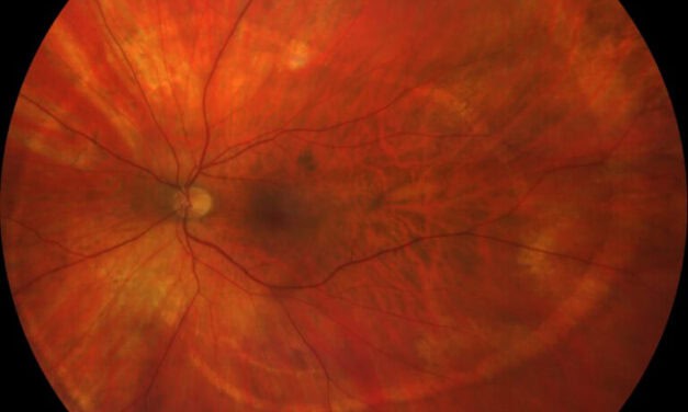 Save as PDF
This case describes an unusual spiral pattern of Schlaegel lines in a patient with inactive multifocal choroiditis. Although most often associated with…
Read More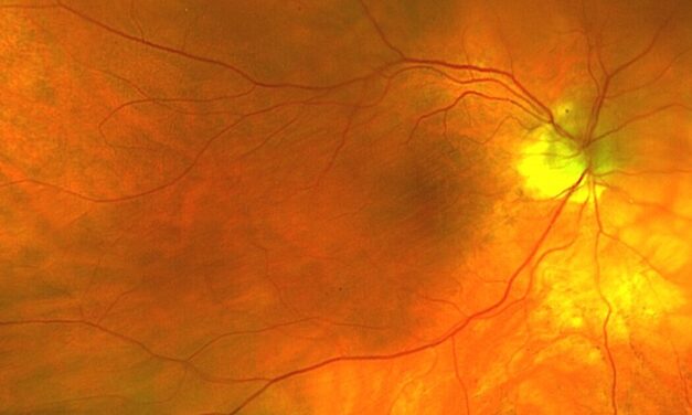 Save as PDF
Choroidal folds are a key indicator of ocular or systemic pathology, with their typical horizontal orientation reflecting underlying anatomical and…
Read More Save as PDF
Peripapillary retinoschisis (PPRS) can cause fluctuations in retinal nerve fiber layer thickness; thus, the spontaneous resolution of PPRS can mimic…
Read More Save as PDF
Babesiosis can cause hemolytic anemia through both infectious and autoimmune processes. Anemic retinopathy can occur due to Babesiosis and…
Read More Save as PDF
PXE is a critical multisystem disease that often presents with readily visible dermatologic manifestations and clinically typical fundus exam findings…
Read More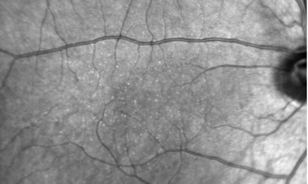 Save as PDF
This case of Benign Yellow Dot Maculopathy represents a unique phenotype that has limited coverage in existing literature. To date, only 48 cases…
Read More Save as PDF
A 49-year-old white man presented to the eye clinic due to sudden-onset scotoma in the right eye. The scotoma was inferior to fixation. He noticed it…
Read More Save as PDF
Differentiation between ischemic and non-ischemic central retinal vein occlusions provides valuable information regarding visual prognosis and risk of…
Read More Save as PDF
Ocular hypotony can occur for a variety of reasons, including but not limited to ocular inflammation, glaucoma filtration surgery, or trauma…
Read More Save as PDF
Waldenstrom macroglobulinemia (WM) is an uncommon lymphoproliferative B-cell disorder characterized by overproduction of monoclonal…
Read More Save as PDF
Multimodal imaging and a thorough case history can aid in the diagnosis of solar maculopathy. OCT and fundus autofluorescence can identify small…
Read More Save as PDF
While patients with RAMs are often asymptomatic, providers must be able to clinically differentiate them from other retinal conditions so they can manage and counsel patients appropriately. This case report reviews the anatomical features, risk factors, clinical course, pathophysiology, and treatment options for potential sequelae of a RAM.
Read More Save as PDF
Macular neovascularization (MNV) is a pathophysiological precursor to local tissue destruction. Age-related macular degeneration is the predominant disease for MNV incidence. The spectrum of macular neovascular morphologies can be studied with optical coherence tomography angiography (OCTA) imaging and observed for vision-threatening activity.
Read More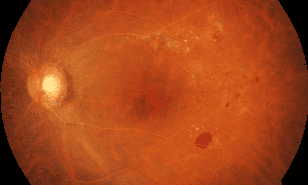 Save as PDF
Severe hypertriglyceridemia can transiently cause the appearance of a salmon-colored fundus with creamy vessels which is known as lipemia retinalis
Read More Save as PDF
MacTel type 2 is the most common subtype that results in atrophic changes and sometimes subretinal neovascularization (SRN). This case report will focus on a patient with MacTel type 2 and the management and treatment options pertinent to the case.
Read More Save as PDF
Torpedo maculopathy is a rare condition resulting in malformation of the outer retina. These lesions are often asymptomatic and found incidentally during routine eye examination. Although typically benign, there are rare reports of associated complications requiring treatment. Appropriate monitoring is thus required.
Read More