

A Blow to the Eye: Managing Traumatic Eye Disease from Commotio Retinae to Macular Hole: A Case Report
 Save as PDF
Conflict of interest: The author(s) declared that this work was conducted in the absence of any commercial or financial relationships that could be construed as a potential conflict of interest.
Abstract
Background: Commotio retinae is a retinal disorder caused by ocular injury, most commonly blunt force trauma to the head or orbit. This condition is characterized by retinal whitening in a coup or contrecoup presentation (translated to “blow” or “counter blow”) where there is a dual-site injury at the initial location and directly opposite to it. Patients with this condition may present asymptomatically if only the peripheral retina is affected, but they may experience blurred vision, metamorphopsias, or scotomas if the macula is involved. This case report will review commotio retinae in addition to potential ocular complications secondary to a blunt force trauma.
Case Report: A 39-year-old African-American male presented with complaints of blurred vision, pain, light sensitivity, and redness in both eyes for one day. He had a history of sustaining an impact from a soccer ball to the right side of his face the previous night. Visual acuity was light perception in the right eye, and 20/20 in the left eye. Confrontation fields were restricted inferiorly in the right eye. Ocular examination revealed a traumatic iritis, vitreous hemorrhage, and pre-retinal hemorrhages in the right eye. Close monitoring for resolution revealed a secondary macular hole two weeks after injury. The patient was referred to a vitreoretinal specialist for further management.
Conclusion: Blunt force trauma to the eye can cause a wide range of ocular complications. Although commotio retinae, traumatic iritis, and hyphema often resolve fully with proper management, more serious complications such as retinal tears, macular holes, and retinal detachment can result in permanent visual consequences. Therefore, a thorough dilated fundoscopic examination is essential. Ancillary testing such as gonioscopy and optical coherence tomography is often necessary to fully assess traumatic sequelae. Eye care practitioners should advocate for safe sports training and educate patients on the risks of sports-related ocular trauma.
Keywords: blunt trauma, commotio retinae, macular hole, optical coherence tomography, traumatic iritis
Introduction
Commotio retinae is a retinal condition resulting from ocular injury, most commonly due to blunt trauma. It is caused by a loss or disruption of the photoreceptor outer segments, which leads to a grey or whitened appearance of the fundus. Commotio retinae most commonly affects the temporal retina because trauma typically affects the nasal globe, sending contrecoup waves temporally.1 The prognosis in mild cases is good, with spontaneous resolution generally within six weeks depending on the severity of the trauma.1 Eyes with severe involvement may have residual retinal pigment epithelium (RPE) changes, RPE atrophy or hyperpigmentation, intraretinal hemorrhage, progressive pigmentary degeneration, retinal detachment, and/or macular hole formation.2 This paper explores clinical features and insights to assist clinicians with the diagnosis and management of commotio retinae and its sequelae.
Case Report
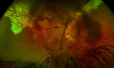
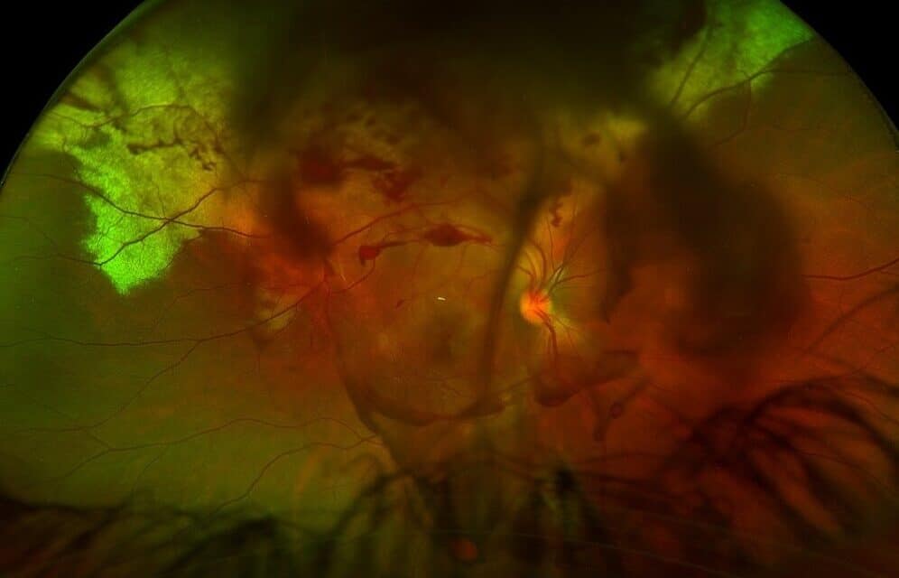
A 39-year-old African-American male presented with sudden, acute loss of vision in his right eye following blunt force trauma involving a soccer ball to the right eye the day before. He reported a cloud of blurred vision that moved with eye movement. His right eye was burning, light sensitive, painful (8 out of 10), and tender to touch. Intraocular pressures were 10 mmHg in the right eye, and 12 mmHg in the left eye. The left eye was also mildly light sensitive. The patient was using over-the-counter Clear Eyes and Tylenol, which offered little relief. He did not report having any flashes or floaters. Corrected visual acuity was light perception in the right eye, and 20/20 in the left eye. Previous ocular and medical history were unremarkable. Pupil assessment revealed mydriasis of the right eye. Both pupils were round and reactive to light and without APD. Extraocular movements were grossly full, although the patient reported discomfort from ocular pain. Confrontation fields were restricted in the inferior quadrants in the right eye. No restriction of the left visual field was noted. Slit lamp biomicroscopy revealed 2+ bulbar conjunctival injection and 4+ cells in the anterior chamber of the right eye. Seidel’s sign was negative, and there was no hyphema, anterior synechiae, nor posterior synechiae in either eye. The anterior segment of the left eye showed mild conjunctival injection and a quiet anterior chamber. A dilated fundus examination of the right eye revealed superior retinal whitening extending to the superior arcades, consistent with the appearance of commotio retinae. A vitreous hemorrhage and multiple pre-retinal hemorrhages in the superior and inferior peripheral retina were also present in this eye (Figure 1). No signs of optic nerve cupping, pallor, or swelling were noted in either eye.
The patient was prescribed prednisolone acetate 1% every two hours and atropine 1% dosed twice a day in the right eye. He was advised to use sunglasses due to the mydriatic side effects of atropine. An urgent referral was placed to a retinal specialist for evaluation with B-scan to rule out a retinal tear or detachment. He was also advised to return to the clinic sooner if any symptoms worsened, such as a decrease in vision, increase in pain, redness or light sensitivity.
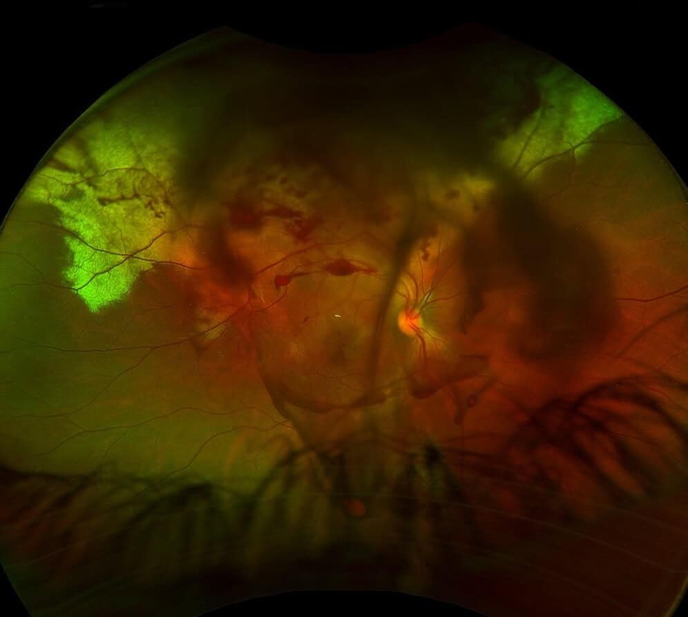
Figure 1. Color fundus image of the right eye at initial presentation. There is a large area of commotio retinae superiorly with adjacent pre-retinal hemorrhages and vitreous hemorrhage surrounding the posterior pole.
Two days following the initial visit, uncorrected visual acuities were counting fingers at 3 feet in the right eye (pinhole 20/200) and 20/20 in the left eye. Intraocular pressures were 12 mmHg OD, and 11 mmHg OS. The patient’s right eye was pharmacologically dilated from the atropine previously prescribed. Upon slit lamp examination, 2+ cells were noted in the right eye and 1+ cell in the left. The cells in the left eye likely appeared a few days after the initial trauma because the injury triggered a slow progressive inflammatory response rather than an immediate one, as seen in the right eye. Retinal findings were stable in the left eye. The right eye had a large area of retinal whitening superiorly, improvement of the pre-retinal hemorrhages, and an expansion of the vitreous hemorrhage (Figure 2). The patient was instructed to continue prednisolone acetate 1% four times a day and atropine 1% twice a day in the right eye and initiate the same regimen in the left eye.
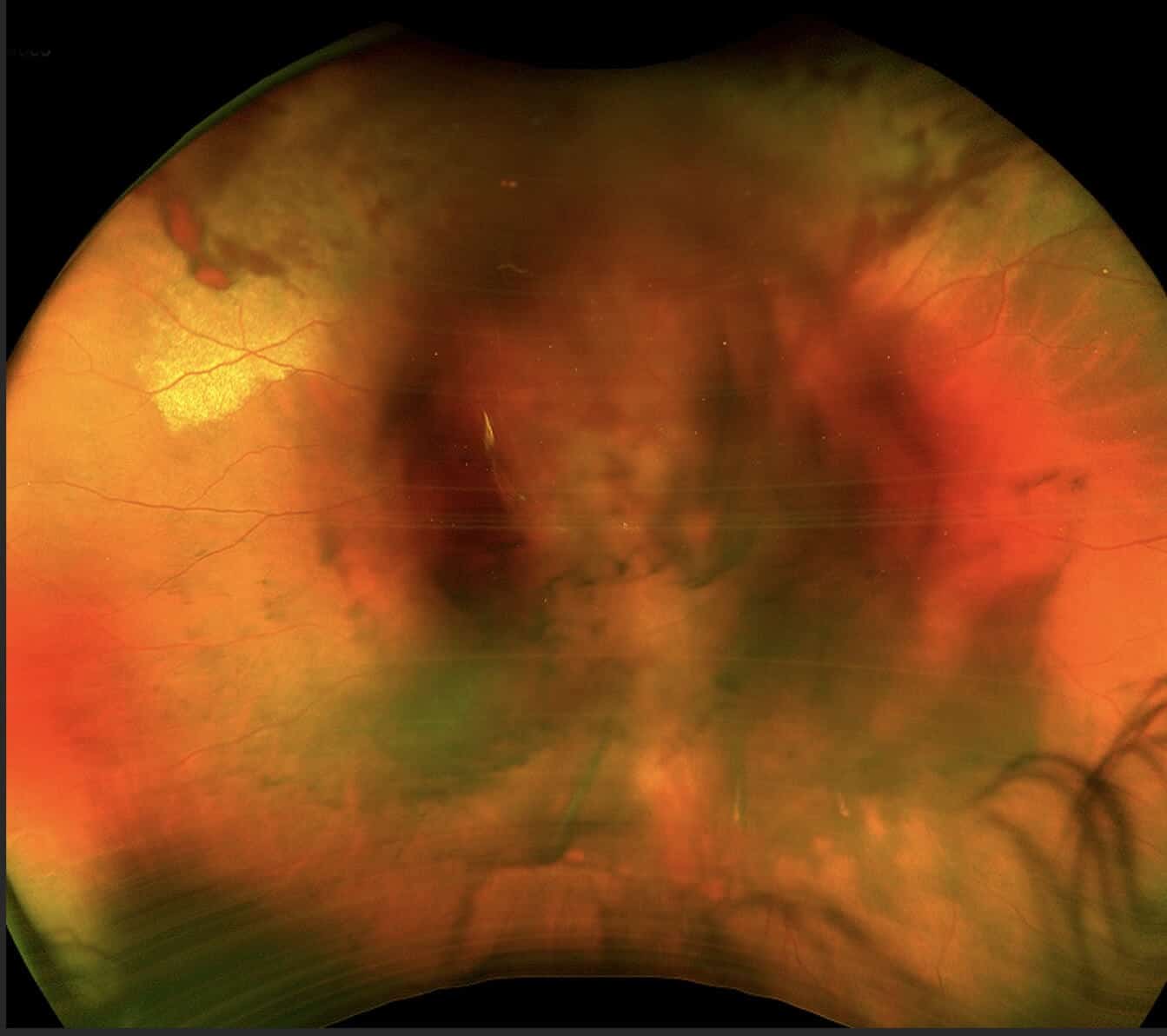
Figure 2. Color fundus image of the right eye three days after the injury. There is stable commotio retinae superiorly with adjacent pre-retinal hemorrhages. The vitreous hemorrhage appears to be more diffuse than previously.
Five days post-trauma, his uncorrected visual acuity was counting fingers at 7 feet in the right eye (pinhole 20/200) and 20/30 in the left eye. Intraocular pressures were 12 mmHg OD, and 11 mmHg OS. No cells or flare were noted in either eye. The vitreous hemorrhage in the right eye was improving, which allowed for clearer retinal views. The commotio retinae was still present but improving throughout the superior and inferior peripheral retina and posterior pole. Out of an abundance of caution with a possible delayed inflammatory response, the patient was advised to maintain his drop dosage to prednisolone acetate 1% at four times a day in both eyes and atropine 1% twice a day in both eyes.
Ten days following the injury, his uncorrected visual acuity had improved to 20/400 and was stable at 20/30 in left eye. The anterior chambers were quiet in both eyes. The patient was pharmacologically dilated from atropine in both eyes. Retina findings were stable in both eyes. The prednisolone acetate 1% taper was started (TID x 3 days, then BID x 3 days, then QD x 2 days) and the atropine 1% was discontinued.
The patient was initially examined by a retina specialist 14 days following injury. A B-scan was not performed; however, the retina specialist documented the absence of retinal tears or detachments over 360 degrees of the peripheral retina in both eyes. Gonioscopy was performed at this visit and showed open angles to the ciliary body, mild trabecular meshwork pigmentation, and no angle recession.
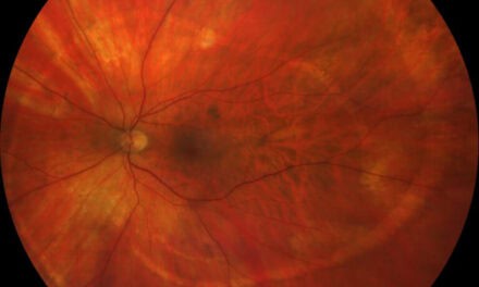
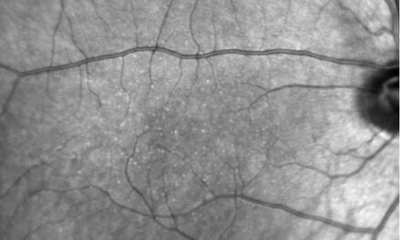
At the next follow up visit, 17 days from the initial incident, the patient still had complaints of persistent blurred vision and light sensitivity. Uncorrected visual acuities were 20/400 in the right eye and 20/20 in the left eye. The anterior chambers remained quiet in both eyes. The pre-retinal hemorrhages and vitreous hemorrhage were resolving, but a new stage IV macular hole measuring approximately 450 µm was noted in the right eye (Figure 3 and 4). He was referred back to the retina specialist within the month for evaluation and consideration of surgical repair.
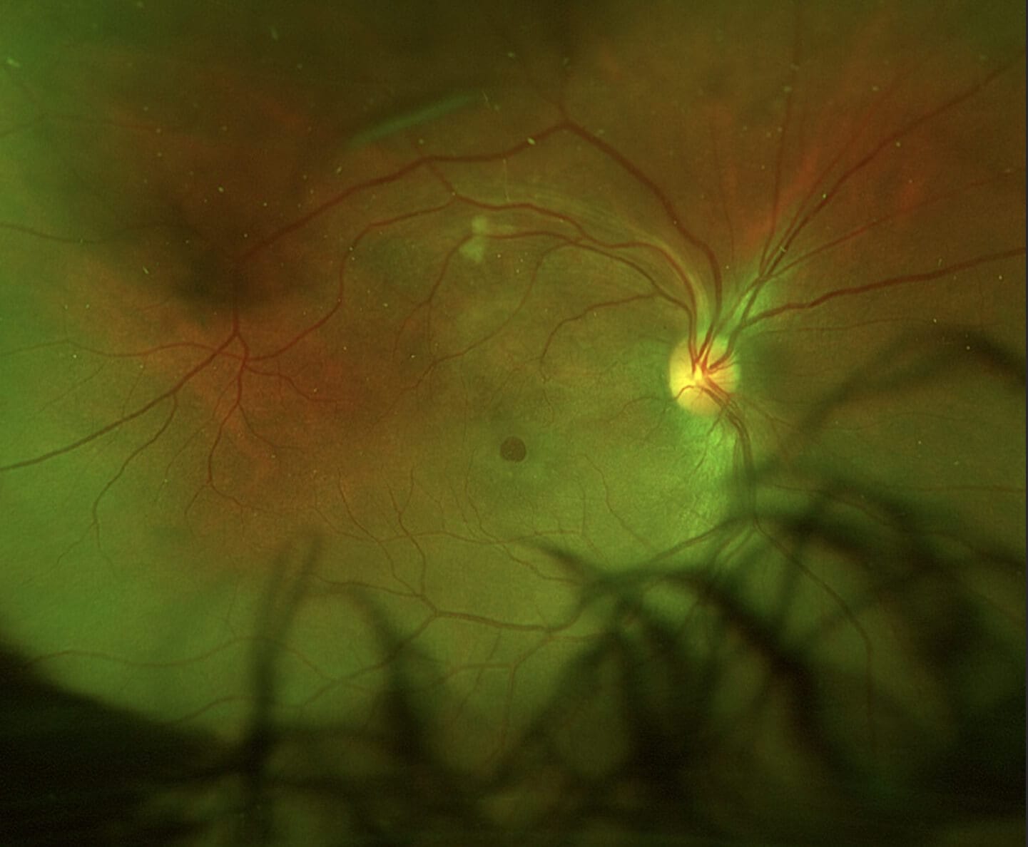
Figure 3. Color fundus image of the right eye showing resolved commotio retina and nearly resolved pre-retinal and vitreous hemorrhages 17 days post-trauma. A newly developed macular hole is also noted.
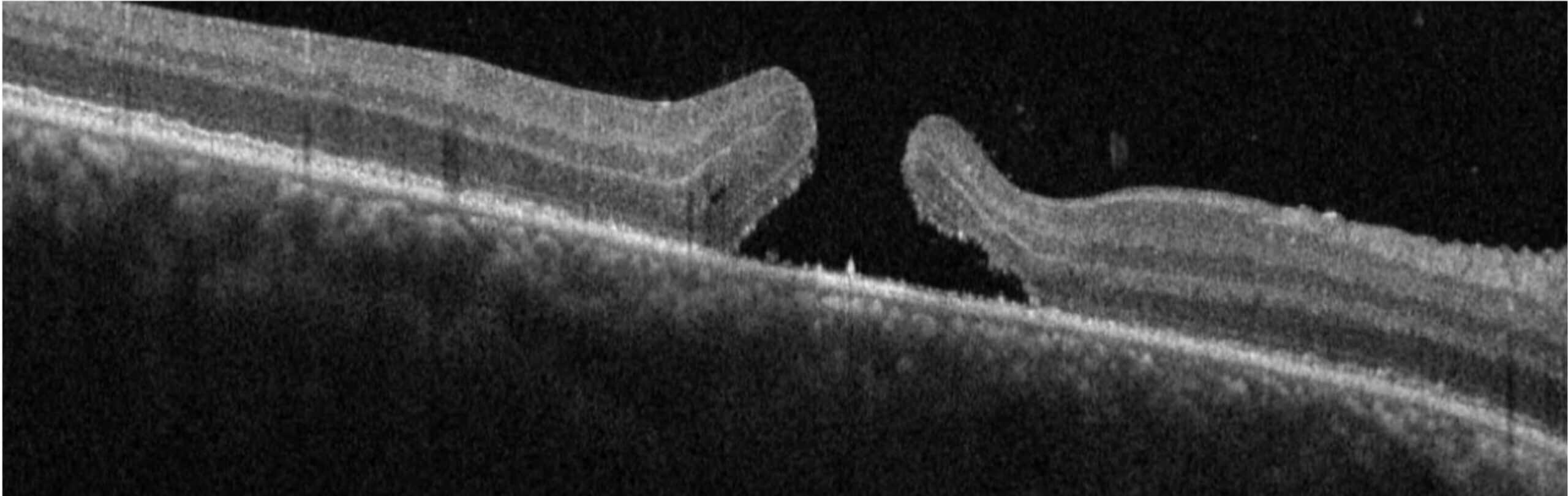
Figure 4. Optical coherence tomography imaging of the initial presentation of a macular hole in the right eye at the two-week follow up.
The patient was seen twice by the retina specialist for evaluation of the hole after its initial presentation, and he was monitored for approximately two months following diagnosis. However, the patient eventually agreed to surgical intervention to repair the macular hole and underwent a pars plana vitrectomy macular hole repair. At the one-month post-operative visit, his vision in the right eye was 20/150 and left eye was 20/20. He was subsequently scheduled for a three-month follow-up visit to monitor.
Discussion
Commotio Retinae
Commotio retinae develops after a traumatic incident to the eye or orbit. Severe blunt trauma to the globe results in anteroposterior compression with simultaneous expansion in the equatorial plane associated with a transient but severe increase in intraocular pressure. Although the impact is primarily absorbed by the lens-iris diaphragm and the vitreous base, damage can also occur at a distant site, such as the retina. Most investigations on contrecoup ocular trauma have been limited to the retina.3 The extent of ocular damage depends on the severity of the trauma and tends largely to be concentrated to either the anterior or posterior segment. Upon fundoscopic examination, retinal whitening is an indicator of trauma to the eye and damage to the retina, which usually resolves within 4-7 days. Disruption of the photoreceptor outer segments is the primary underlying pathology with sparing of the retinal blood vessels.4
While several theories exist for the cause of retinal whitening, a recent thought is that edema is a result of intracellular damage to the Mueller’s cells and mitochondrial swelling in the nerve fiber layer.5 In animal models, the pathogenesis of commotio retinae has included extracellular edema, intracellular edema, and photoreceptor outer segment disruption. Histopathologic findings obtained within 24 hours of blunt trauma in a human eye with clinically observed commotio retinae revealed photoreceptor outer segment disruption and damage to the retinal pigment epithelium, with the major site of injury at the level of the photoreceptor outer segments and retinal pigment epithelium junction.6
Blunt ocular trauma stretches the retina via vitreoretinal attachments at the level of the photoreceptor outer segments. The remaining retina is still held together by the processes of Mueller cells. In addition to the role of the deformation of the vitreous (traction and compression of the retina), hydraulic forces generated from blunt trauma are transmitted posteriorly and may result in mechanical disruption of the photoreceptor outer segments.7
The most common causes of ocular trauma are associations with high impact activities such as squash balls, elastic luggage straps, and champagne corks. Ocular trauma is the main cause of unilateral vision loss in male patients between 22 and 44 years of age.8 The prevalence of commotio retinae has been estimated to be 30 to 90% of blunt force traumas.9 There is also a higher risk with assault-related incidences as compared to those falling after standing up.10
The patient’s symptoms are dependent on the location of the retinal injury. Most peripheral retinal injuries are asymptomatic. However, if the posterior pole is involved with commotio retinae, the edema is referred to as Berlin’s edema, and patients will present with symptoms such as blurred vision, scotoma, and, possibly, changes in color vision. Commotio retinae can present with a “pseudo cherry red spot” at the macula as a result of peripheral retinal whitening.
The most common retinal locations of commotio retinae occur inferotemporal (37%), temporal (17%), and supratemporal (17%). These locations directly correlate with the likelihood of injury due to their proximity to the edge of the face.11 However, in patients with orbital wall fractures, most commonly medial or inferior wall fractures, more than half of the commotio retinae cases were found to be inferiorly located.10
In general, ocular trauma commonly results in long-term effects; the prognosis is therefore guarded. However, in commotio retinae specifically, most patients recover in 1-4 weeks without permanent defects.4 Although a retrospective study reported that after an injury leading to macular damage, 74% of patients’ vision recovered to greater than or equal to 20/30.1 This can still be significant to patients due to symptomatic paracentral visual defects and high patient awareness of a reduction of vision from 20/15 to 20/30.1 Another retrospective study reported an injury with an orbital wall fracture had resolution of 20/25 or better.10
Optical coherence tomography (OCT) imaging is a helpful non-invasive tool to assist with monitoring retinal changes. OCT findings can range from mild lesions with transient hyper-reflectivity of the ellipsoid layer of the outer retina as well as a loss of the thin hypo-reflective optical space, to more severe cases presenting with a disruption in the inner and outer segment junction, hyper-reflectivity of the overlying retina, pigment disorders, and retinal atrophy.8,12 While the appearance is one of intracellular edema, if the diagnostic imaging with macular OCT shows no increase in retinal thickness, this indicates the cloudiness is a disruption of photoreceptor cells rather than fluid buildup.
| Grade | Optical coherence tomography feature |
|---|---|
| 1 | Increase in inner segment/outer segment (IS-OS) junction reflectivity |
| 2 | Loss of reflection in cone outer segment tips (COST) |
| 3 | COST and IS/OS junction defects |
| 4 | COST, IS/OS junction, and external limiting membrane (ELM) defects |
Another ancillary test used for commotio retinae is fluorescein angiography (FA) and fundus autofluorescence (FAF). FA and FAF imaging can be useful in detecting early retinal changes by focusing on the fluorescent properties of retinal pigment that are not as easily detected with a standard dilated fundus examination.14 There is no evidence of alteration of retinal vasculature or choroidal permeability in commotio retinae; however, hyper-fluorescent areas from a window defect or hypo-fluorescent areas from a blockage of blood or another opacity can block the fluorescence in these images. Once the retinal whitening has reversed, the areas return to normal.8
Hyphema
In most hyphema cases, there is resolution without medical intervention; however, a traumatic hyphema may be associated with intraocular pressure (IOP) elevation due to trabecular blockage by red blood cells. Although most traumatic hyphemas are relatively innocuous and transient, severe and prolonged elevation of IOP may damage the optic nerve or cause blood staining of the cornea.
Angle Recession
Angle recession involves rupture of the ciliary body face, the portion of the ciliary body that lies between the iris root and the scleral spur, due to blunt trauma. The rise in IOP is secondary to associated damage to the trabecular meshwork rather than from angle recession itself, yet the risk of glaucoma is directly related to the extent of angle recession.15 A sign of angle recession is an irregular widening of the ciliary body band on gonioscopy.
Gonioscopy is not generally required when vitreous hemorrhages do not originate from proliferative causes, such as from a posterior vitreous detachment, retinal tear, or retinal detachment. However, gonioscopy should be considered after clearing of a traumatic vitreous hemorrhage or hyphema from trauma to rule out angle recession. Since glaucoma may not develop until months or years post-injury, angle recession warrants periodic review.16
Traumatic Iritis
Traumatic iritis may occur after blunt trauma or post-surgically. This form of uveitis is from the breakdown of the blood-aqueous barrier, which is typically self-limiting in nature.17 Symptoms of posttraumatic iritis include blurred vision, throbbing pain, lacrimation, redness, and photophobia.18 Turbidity of the aqueous from white blood cell infiltration and protein exudation results in the blurring of vision.17 Varying degrees of pain seen in anterior uveitis can be attributed to ciliary muscle spasm. Severe pain can be associated with raised intraocular pressures.17,18 Photophobia is commonly due to ciliary muscle spasm, yet anterior chamber cellular infiltration, corneal epithelial edema, and pupillary muscle involvement can also contribute.17 Inflammation can produce either raised or lowered intraocular pressure. Lower intraocular pressure is typically associated with lower inflammation, which causes ciliary body hyposecretion, while higher intraocular pressure is associated with a decrease in aqueous flow as a result of inflammatory cells obstructing the outflow of aqueous from the trabecular meshwork. Corticosteroids are the drug of choice in the treatment of anterior uveitis. Steroids act by modifying and decreasing the inflammatory ocular response. If dosed frequently, topical corticosteroids can reach adequate therapeutic levels in the anterior chamber. Mydriatics serve as supportive therapy and cause paresis of the iris and ciliary muscle.17 These pharmacological agents prevent posterior synechiae and help stabilize the blood-aqueous barrier, thereby preventing additional protein leakage and flare.
Vitreous Hemorrhage
A vitreous hemorrhage is defined as the presence of extravasated blood in the anteriorly in the vitreous cavity between the posterior lens capsule and zonules of the lens, and laterally and posteriorly between the non-pigmented epithelium of ciliary body and internal limiting membrane (ILM).19,20 Trauma is the most common cause of vitreous hemorrhage in patients less than 40 years of age with a male preponderance.21 Trauma may indirectly lead to vitreous hemorrhage because of acute posterior vitreous detachment (PVD) with possible secondary retinal tears.22 The risk of development of retinal tears may be as high as 70% in patients with acute PVD with vitreous hemorrhage.20
B-scan ultrasonography is a key tool for identifying underlying retinal pathology that a hemorrhage could obstruct peripheral funduscopic examination. A detached retina is usually still attached to the anchoring points of the ora serrata and the optic nerve head; consequently, on B-scan, a totally detached retina has a funnel shape.19 While B-scan ultrasonography is an important part of the workup for vitreous hemorrhage patients, it’s not a very sensitive tool. In a pool of 71 patients, Foo et al. found B-scan testing had a 11.53% sensitivity for retinal tears, and 17% sensitivity for discovering rhegmatogenous retinal detachments.23 For fundus-obscuring nondiabetic vitreous hemorrhages, repeat imaging every two weeks is necessary until retinal views are unobstructed, as retinal detachments can occur even if one was not discovered at the initial B-scan.23
In an otherwise healthy patient, acute vitreous hemorrhages may clear over time without intervention. Nonetheless, waiting for the hemorrhage to clear comes with a concern that the hemorrhage is obscuring a complication requiring surgical intervention, particularly in the peripheral retina. Observation and surgical intervention have remained the chief management options for vitreous hemorrhage. The definitive treatment for vitreous hemorrhage is a pars plana vitrectomy, which is commonly indicated in patients with dense, non-clearing vitreous opacities. In general, with the absence of retinal tear, a vitrectomy may be indicated within 2-4 weeks for a non-clearing vitreous hemorrhage.24 Significant improvements in best corrected visual acuity are noted in vitrectomy-treated groups, compared to those without surgical intervention.23,25
Macular Hole
Macular holes can develop after blunt trauma, leaving vision between 20/30 and 20/400, depending on the severity. Full-thickness macular holes are defined as greater than 400 microns in diameter with a red base in which yellow-white dots may be seen. A surrounding grey cuff of subretinal fluid is usually present, and an overlying operculum (sometimes called a pseudo-operculum) may be visible. Visual acuity is often reduced to 20/200, although it is occasionally better, particularly in patients able to use eccentric fixation.26,27
While idiopathic macular holes can take months or even years to form, traumatic macular holes typically form immediately after. However, there are reports indicating a slower formation in the weeks following the injury.27 Two primary theories explain this phenomenon. The tangential vitreoretinal traction theory suggests axial blunt force trauma causes an equatorial expansion, transmitting force to the macula and resulting in a central vision defect.27 Alternatively, the anteroposterior vitreoretinal theory proposes that direct traction on the fovea leads to foveal tissue loss. However, this theory is less likely, as evidence shows a hole can develop a few days post-trauma, then spontaneously close a month later after the vitreous detaches from the fovea.26,27
While most macular holes in adults are idiopathic, the most common etiology in children is trauma. They do, however, have a high spontaneous closure rate between 10% and 57% in the first four months.26 The incidence of traumatic macular holes is 1.4% in closed-globe trauma, and much lower in open-globe injuries.27
The Watzke-Allen Test can help diagnose a full-thickness macular hole, whether idiopathic or traumatic.2,28 It is performed by projecting a narrow slit beam over the center of the hole both vertically and horizontally. A patient with a macular hole will report that the beam is thinned or broken.
OCT imaging is the primary method used to assist with diagnosing macular holes. Gass’s classification of idiopathic macular holes has been revised by Huang et al to specify traumatic macular holes based on OCT imaging as listed below (Table 2):
| Type | Description |
|---|---|
| I | Cystic edema of the neurosensory retina on both margins of the hole on both horizontal and vertical scans |
| II | Cystic edema of the neurosensory retina on only one margin of the hole on either horizontal or vertical scans |
| III | Full thickness defect of neurosensory retina without cystic edema or detachment of margins |
| IV | Localized detachment of the neurosensory retina at the margin without cystic edema |
| V | Thinning of the neurosensory retina |
Based on this grading system, the findings in this case are most congruous with a Type III macular hole 17 days post-trauma.29
The management of traumatic macular holes is similar to that of idiopathic macular holes. Most are monitored in the acute stage, as there is a 50% spontaneous closure rate in children and a 28.6% rate in adults following a traumatic macular hole.27 There is a window of 5.6 weeks of spontaneous closure, but anything beyond 67.3 weeks did not yield spontaneous closure.27 Macular hole closure is generally associated with visual acuity improvement of at least two lines. The most likely holes with spontaneous closure were those that did not have intraretinal cysts and had a small minimum linear diameter.30 Those with intraretinal cysts were prescribed nonsteroidal anti-inflammatory drugs, namely diclofenac and ketorolac, to decrease the edema, increasing the likelihood of hole closure.31 A theory on why this happens is that glial cells proliferate from the retinal pigment epithelium to the basal surface of the hole, promoting closure.26,27
Median symptom duration was 6 months with primary closure achieved in 81.5% of eyes demonstrating a linear relationship between predicted probability of closure and symptom duration.32 Many reports describe spontaneous closure within two months of trauma. Waiting one to three months before intervening with surgery seems prudent. Johnson et al. alluded to Berlin’s edema being associated with photoreceptor and RPE damage and mentioned 4 eyes that had significant RPE changes after surgery with final visual outcome ranging from 20/30 to 20/200.33
It is recommended to have an internal limiting membrane (ILM) peel within six months for best visual recovery.34 Traumatic holes that were delayed to have a vitrectomy after a year or more were less likely to close.35 Surgical intervention of macular holes as originally described by Wendel et al. follows the following five steps: 1) pars plana vitrectomy 2) induction of a posterior vitreous detachment, if not present 3) epiretinal membrane peel 4) fluid-gas exchange 5) one week of occiput-up positioning.36 However, Kuhn et al. were among the first to report success in peeling the ILM prior to fluid-air exchange, specifically in traumatic macular holes.37 This use of a gas tamponade agent, such as SF6 or C3F8, has reported increased success rates of 90 to 92.3% as compared to a silicone oil agent at 67 to 90%.38
Conclusion
Commotio retinae occurs as a result of trauma to the eye with a generally good prognosis; however, in the case of macular holes, closer management is needed. Spontaneous closure of the macular hole is common within the first 3 to 6 months, so immediate surgical intervention is not always necessary. Eye care professionals can help provide the best visual outcome for these patients by obtaining a careful history, performing a thorough dilated exam, and in utilizing special testing for the evaluation of the retina, such as OCT and B-scan. Patient education and compliance are also crucial to ensure the best visual prognosis for these patients.
References
- Blanch RJ, Good PA, Shah P, Bishop JRB, Logan A, Scott RAH. Visual outcomes after blunt ocular trauma. Ophthalmology. 2013 Aug;120(8):1588–91. doi:10.1016/j.ophtha.2013.01.009 PubMed PMID: 23618228.
- Salmon JF, Bowling B. Kanski’s clinical ophthalmology : a systematic approach. Ninth edition. Edinburgh: Elsevier; 2020. (Clinical ophthalmology).
- Wolter JR. Coup-contrecoup mechanism of ocular injuries. Am J Ophthalmol. 1963 Nov;56:785–96. doi:10.1016/0002-9394(63)92943-x PubMed PMID: 14077183.
- Sharief S, Jayadev C, Gadde SGK. Commotio retinae halo. BMJ Case Rep. 2022 Mar 17;15(3). doi:10.1136/bcr-2022-249270 PubMed PMID: 35301188; PubMed Central PMCID: PMC8932277.
- Bunt-Milam AH, Black RA, Bensinger RE. Breakdown of the outer blood-retinal barrier in experimental Commotio retinae. Exp Eye Res. 1986 Sep 1;43(3):397–412. doi:10.1016/S0014-4835(86)80076-8
- Mansour AM, Green WR, Hogge C. Histopathology of commotio retinae. Retina Phila Pa. 1992;12(1):24–8. doi:10.1097/00006982-199212010-00006 PubMed PMID: 1565867.
- Mohseni M, Gurnani B, Blair K. Blunt Eye Trauma. In: StatPearls. Treasure Island (FL): StatPearls Publishing; 2026. PubMed PMID: 29261988.
- Mendes S, Campos A, Campos J, Neves A, Beselga D, Fernandes C, et al. Cutting Edge of Traumatic Maculopathy with Spectral-domain Optical Coherence Tomography – A Review. Med Hypothesis Discov Innov Ophthalmol J. 2015 Summer;4(2):56–63. PubMed PMID: 26060831; PubMed Central PMCID: PMC4458327.
- Leshno A, Alhalel A, Fogel-Levin M, Zloto O, Moisseiev J, Vidne-Hay O. Pediatric retinal damage due to soccer-ball-related injury: Results from the last decade. Eur J Ophthalmol. 2021 Jan 1;31(1):240–4. doi:10.1177/1120672119882332
- Blegen HMJ, Santamaria JA, Mehta A, Reed DS, Drayna PM, Davies B. Patterns and prognosis of commotio retinae in orbital wall fractures. Ther Adv Ophthalmol. 2019 Dec;11:2515841419862133. doi:10.1177/2515841419862133 PubMed PMID: 31321382; PubMed Central PMCID: PMC6628538.
- Venugopal R, Das AV, Takkar B, Stewart MW, Narayanan R. Real-world experience of full-thickness traumatic macular hole among young patients. Int J Retina Vitr. 2024 Feb 21;10(1):20. doi:10.1186/s40942-024-00539-3 PubMed PMID: 38383490; PubMed Central PMCID: PMC10882818.
- Venkatesh R, Pereira A, Sangai S, Yadav NK. Hyporeflective micro-elevations and irregularity of the ellipsoid layer: novel optical coherence tomography features in commotio retinae. Can J Ophthalmol J Can Ophtalmol. 2020 Dec;55(6):492–9. doi:10.1016/j.jcjo.2020.06.020 PubMed PMID: 32853587.
- Ahn SJ, Woo SJ, Kim KE, Jo DH, Ahn J, Park KH. Optical coherence tomography morphologic grading of macular commotio retinae and its association with anatomic and visual outcomes. Am J Ophthalmol. 2013 Nov;156(5):994-1001.e1. doi:10.1016/j.ajo.2013.06.023 PubMed PMID: 23972302.
- Sepah YJ, Akhtar A, Sadiq MA, Hafeez Y, Nasir H, Perez B, et al. Fundus autofluorescence imaging: Fundamentals and clinical relevance. Saudi J Ophthalmol Off J Saudi Ophthalmol Soc. 2014 Apr;28(2):111–6. doi:10.1016/j.sjopt.2014.03.008 PubMed PMID: 24843303; PubMed Central PMCID: PMC4023118.
- Herschler J. Trabecular damage due to blunt anterior segment injury and its relationship to traumatic glaucoma. Trans Sect Ophthalmol Am Acad Ophthalmol Otolaryngol. 1977 Apr;83(2):239–48. PubMed PMID: 406709.
- Kaufman JH, Tolpin DW. Glaucoma after traumatic angle recession. A ten-year prospective study. Am J Ophthalmol. 1974 Oct;78(4):648–54. doi:10.1016/s0002-9394(14)76303-2 PubMed PMID: 4413576.
- Agrawal RV, Murthy S, Sangwan V, Biswas J. Current approach in diagnosis and management of anterior uveitis. Indian J Ophthalmol. 2010 Feb;58(1):11–9. doi:10.4103/0301-4738.58468 PubMed PMID: 20029142; PubMed Central PMCID: PMC2841369.
- Hogan MJ, Kimurs SJ, Thygeson P. Signs and symptoms of uveitis. I. Anterior uveitis. Am J Ophthalmol. 1959 May;47(5 Pt 2):155–70. doi:10.1016/s0002-9394(14)78239-x PubMed PMID: 13649855.
- Yanoff M, Duker J. Ophthalmology. 3rd ed. 2008.
- Wingelaar M, Shah G. Vitreous Hemorrhage: Obeserve or operate. 2022 Oct.
- Jena S, Tripathy K. Vitreous Hemorrhage. In: StatPearls. Treasure Island (FL): StatPearls Publishing; 2026. PubMed PMID: 32644557.
- Byer NE. Natural history of posterior vitreous detachment with early management as the premier line of defense against retinal detachment. Ophthalmology. 1994 Sep;101(9):1503–13; discussion 1513-1514. doi:10.1016/s0161-6420(94)31141-9 PubMed PMID: 8090453.
- Foo E, Grassi P, Spiteri-Cornish K. Early vitrectomy in eyes with non-diabetic vitreous hemorrhage. Ther Adv Ophthalmol. 2022 Dec;14:25158414221090099. doi:10.1177/25158414221090099 PubMed PMID: 35510165; PubMed Central PMCID: PMC9058341.
- Treumer F, Roider J. [Vitreous body hemorrhage-How long can one wait?]. Ophthalmol Z Dtsch Ophthalmol Ges. 2020 Sep;117(9):866–70. doi:10.1007/s00347-020-01112-7 PubMed PMID: 32385553.
- Pighin MS, Berrozpe C, Jürgens I. Outcome of acute nontraumatic vitreous hemorrhage in healthy patients. Retina Phila Pa. 2020 Jan;40(1):87–91. doi:10.1097/IAE.0000000000002338 PubMed PMID: 30300268.
- Kusaka S, Fujikado T, Ikeda T, Tano Y. Spontaneous disappearance of traumatic macular holes in young patients. Am J Ophthalmol. 1997 Jun;123(6):837–9. doi:10.1016/s0002-9394(14)71136-5 PubMed PMID: 9535631.
- Budoff G, Bhagat N, Zarbin MA. Traumatic Macular Hole: Diagnosis, Natural History, and Management. J Ophthalmol. 2019;2019:5837832. doi:10.1155/2019/5837832 PubMed PMID: 31016038; PubMed Central PMCID: PMC6444256.
- Fischer C, Callizo J, Wetzel E, Feltgen N, Hoerauf H. [Importance of the Watzke-Allen test in diagnostics and staging of macular holes]. Ophthalmol Z Dtsch Ophthalmol Ges. 2016 Feb;113(2):152–5. doi:10.1007/s00347-015-0102-y PubMed PMID: 26205743.
- Huang J, Liu X, Wu Z, Lin X, Li M, Dustin L, et al. Classification of full-thickness traumatic macular holes by optical coherence tomography. Retina Phila Pa. 2009 Mar;29(3):340–8. doi:10.1097/IAE.0b013e31819241d0 PubMed PMID: 19092730; PubMed Central PMCID: PMC5340151.
- Chen H, Chen W, Zheng K, Peng K, Xia H, Zhu L. Prediction of spontaneous closure of traumatic macular hole with spectral domain optical coherence tomography. Sci Rep. 2015 Jul 21;5:12343. doi:10.1038/srep12343 PubMed PMID: 26196460; PubMed Central PMCID: PMC4508835.
- Li AS, Ferrone PJ. Traumaic macular hole closure and visual improvement after topical nonsteroidal antiinflammatory drug treatment. Retin Cases Brief Rep. 2020 Fall;14(4):324–7. doi:10.1097/ICB.0000000000000705 PubMed PMID: 29384982.
- Murphy DC, Al-Zubaidy M, Lois N, Scott N, Steel DH. The Effect of Macular Hole Duration on Surgical Outcomes: An Individual Participant Data Study of Randomized Controlled Trials. Ophthalmology. 2023 Feb;130(2):152–63. doi:10.1016/j.ophtha.2022.08.028 PubMed PMID: 36058348.
- Johnson RN, McDonald HR, Lewis H, Grand MG, Murray TG, Mieler WF, et al. Traumatic macular hole: observations, pathogenesis, and results of vitrectomy surgery. Ophthalmology. 2001 May;108(5):853–7. doi:10.1016/s0161-6420(00)00650-3 PubMed PMID: 11320012.
- Shukla SY, Afshar AR, Kiernan DF, Hariprasad SM. Outcomes of chronic macular hole surgical repair. Indian J Ophthalmol. 2014 Jul;62(7):795–8. doi:10.4103/0301-4738.138302 PubMed PMID: 25116773; PubMed Central PMCID: PMC4152650.
- Miller JB, Yonekawa Y, Eliott D, Kim IK, Kim LA, Loewenstein JI, et al. Long-term Follow-up and Outcomes in Traumatic Macular Holes. Am J Ophthalmol. 2015 Dec;160(6):1255-1258.e1. doi:10.1016/j.ajo.2015.09.004 PubMed PMID: 26393438.
- Wendel RT, Patel AC, Kelly NE, Salzano TC, Wells JW, Novack GD. Vitreous surgery for macular holes. Ophthalmology. 1993 Nov;100(11):1671–6. doi:10.1016/s0161-6420(93)31419-3 PubMed PMID: 8233393.
- Kuhn F, Morris R, Mester V, Witherspoon CD. Internal limiting membrane removal for traumatic macular holes. Ophthalmic Surg Lasers. 2001 Aug;32(4):308–15. PubMed PMID: 11475397.
- Ghoraba HH, Ellakwa AF, Ghali AA. Long term result of silicone oil versus gas tamponade in the treatment of traumatic macular holes. Clin Ophthalmol Auckl NZ. 2012;6:49–53. doi:10.2147/OPTH.S22061 PubMed PMID: 22259236; PubMed Central PMCID: PMC3259100.

Dr. Glass earned her optometry degree from the University of Houston College of Optometry. Her training included rotations in ocular disease and pediatric eye exams at La Nueva Casa de Amigos in Houston, Texas, and ocular disease management at Northeastern State University Oklahoma College of Optometry in Tahlequah, Oklahoma. Her focus in optometry has been general family eye care, pediatric eye care, and dry eye management. She is a member of the American Optometric Association, Oklahoma Association of Optometric Physicians, and a lifetime member of Mensa, the International High IQ Society.

Dr. Hecker earned his Doctor of Optometry degree from Northeastern State University Oklahoma College of Optometry in 2020 and completed a residency in ocular disease and primary eye care at the Jack C. Montgomery VA Medical Center. He is currently a staff optometrist at the Ernest Childers VA Outpatient Clinic, where he provides patient care and clinical teaching for students and residents. He also serves as adjunct faculty with several schools of optometry.

Joann Cherian is an optometrist practicing at the Veterans Affairs in Oklahoma City, where she is dedicated to providing comprehensive eye care to veterans. She earned her Doctorate of Optometry degree from the University of Houston College of Optometry, is a Fellow of the American Academy of Optometry, and is a Diplomate of the American Board of Optometry. Outside of her clinical work, Joann enjoys traveling with her husband, spending time with family and friends, and trying new restaurants.

Dr. Kelly graduated from the University of Houston College of Optometry. She completed an ocular disease/primary care residency at The Veteran’s Health Care System of the Ozarks. She has served on staff there since the completion of her residency and works with students and residents. She enjoys hiking, cycling and traveling with her husband and two daughters.