

A Case of Incidental Diagnosis of Subclinical Sectoral Non-Arteritic Anterior Ischemic Optic Neuropathy in a Glaucoma Suspect
 Save as PDF
Conflict of interest: The author(s) declared that this work was conducted in the absence of any commercial or financial relationships that could be construed as a potential conflict of interest.
Abstract
Background: Non-arteritic anterior ischemic optic neuropathy (NAION) and glaucoma are the two most prevalent optic neuropathies associated with retinal ganglion cell and axonal loss.1,2 When both conditions coexist, careful distinction between their mechanisms, etiologies, and clinical presentations is essential for accurate diagnosis and management. Although NAION and glaucoma share several vascular and systemic risk factors, they differ in pathophysiology and clinical course. Subclinical, sectoral NAION incidentally detected on OCT (Optical Coherence Tomography) in an asymptomatic glaucoma suspect without clinically evident optic disc edema is uncommon and presents a diagnostic challenge. This case highlights the importance of correlating OCT findings with functional testing and longitudinal follow-up to determine the primary etiology of axonal loss and to guide appropriate clinical decision-making.
Case Report: A 50-year-old African American male presented for a routine eye examination. He was followed as a glaucoma suspect of both eyes, with greater concern in the left eye, due to a history of ocular trauma with possible angle recession in the left eye. Best-corrected visual acuity was 20/20 in each eye with spectacles, and the patient had normal color vision, pupils, extraocular motility, and confrontation fields. Dilated fundus examination showed healthy optic nerves without edema bilaterally.
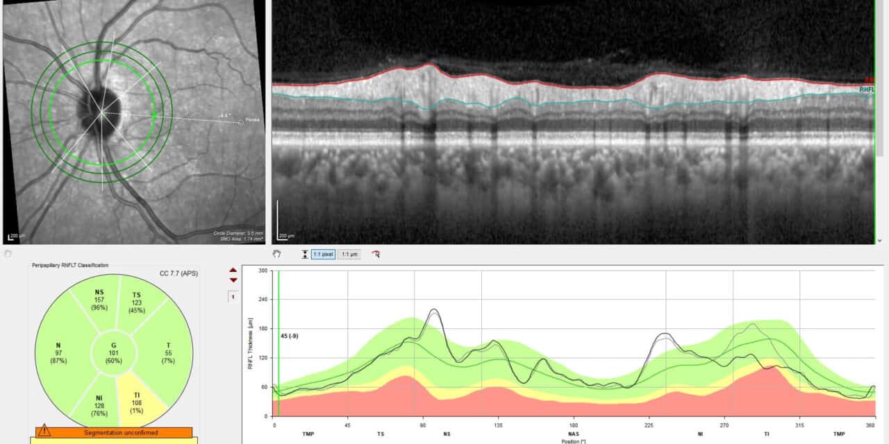
Optical Coherence Tomography demonstrated a nearly 50µm inferior temporal sectoral thickening of the retinal nerve fiber layer (RNFL) in the left eye, while the right eye remained stable. Humphrey visual field (HVF) testing revealed a corresponding superior nasal defect in the left eye. The findings were considered most consistent with suspected incipient subclinical sectoral NAION of the left eye. Later OCTs demonstrated a nearly 50µm inferior temporal sectoral thinning of the RNFL in the left eye from baseline over 10 months, which might have been attributed to glaucoma if he had not incidentally presented for his routine exam at this particular time.
Conclusion: This case highlights incidentally detected subclinical sectoral NAION in a glaucoma suspect and the diagnostic challenge of differentiating ischemic from glaucomatous optic neuropathy. The transient RNFL thickening detected on OCT was critical in identifying an acute ischemic event that might otherwise have been misinterpreted as glaucomatous progression at a later time. Had this patient been evaluated at a different time point, the residual RNFL thinning and field defect could have reasonably prompted a diagnosis of glaucomatous optic neuropathy with subsequent treatment initiation. Given similar vascular risk factors and similarities in structural sequelae, careful correlation of clinical findings, imaging, and disease course is essential for accurate diagnosis, avoidance of unnecessary therapy, and appropriate assessment of progression.
Keywords: Non-arteritic anterior ischemic optic neuropathy, glaucoma, optical coherence tomography, Humphrey visual field
Introduction
Non-arteritic anterior ischemic optic neuropathy (NAION) and glaucoma represent the two most common causes of acquired optic nerve-related visual field loss in adults, characterized by retinal ganglion cell degeneration.1,2 Despite their shared consequence of irreversible axonal loss, the disorders differ in their pathophysiology. NAION results from acute hypoperfusion of the optic nerve head, whereas glaucoma represents a chronic, multifactorial injury to ganglion cells.1,2,3 Distinction between the conditions relies on clinical optic nerve appearance, visual field loss, and structural imaging. However, these features may overlap once an ischemic event, such as NAION, has resolved.4,5 The challenge is further heightened when an acute NAION is sectoral, and the optic nerve appears to have distinct disc margins without swelling or hemorrhaging upon initial clinical examination. Accurate differentiation is essential to avoid misdiagnosis and unnecessary therapy.
Case Report
A 50-year-old African American male presented as an established patient for a routine eye examination and OCT. He was followed as a glaucoma suspect, greater in the left eye due to prior ocular trauma with possible angle recession. He has had stable OCTs and clear visual fields since baseline eye exam five years prior. Ocular history included pterygium excision of the left eye, dry eye syndrome, a pigmented vitreoretinal tuft of the left eye, myopia, and presbyopia. Medical history included borderline obstructive sleep apnea (OSA) diagnosed eight years prior, PTSD, depression, and anxiety. Vital signs revealed blood pressure of 101/66 mmHg, pulse of 49 bpm, and an average BMI. Current medications included fluoxetine, hydroxyzine, carboxymethylcellulose, and ketotifen.
Best-corrected visual acuity was 20/20 in each eye with spectacles, and the patient had normal color vision, pupils, extraocular motility, and confrontation fields. Slit lamp examination revealed mild meibomian gland dysfunction, nasal corneal surgical scarring status post resection of the pterygium of the left eye, and superficial punctate epithelial erosions of both eyes. Intraocular pressure (IOP) was 21 mmHg in each eye with Goldman Applanation Tonometry at 11:42AM. Dilated fundus examination revealed healthy, flat optic nerves bilaterally with a 0.35 cup/disc ratio of the right eye and 0.25 cup/disc ratio of the left eye. Of note, both nerves were mildly malinserted, and examination revealed pink and healthy neuro-retinal rims with flat and distinct disc margins 360°, strong positive spontaneous venous pulsation, no vessel obscuration, and no clinically apparent edema of either eye. Peripheral retinal exam of the right eye was unremarkable. A pigmented vitreoretinal tuft was noted in the inferior periphery of the left eye without an associated break.
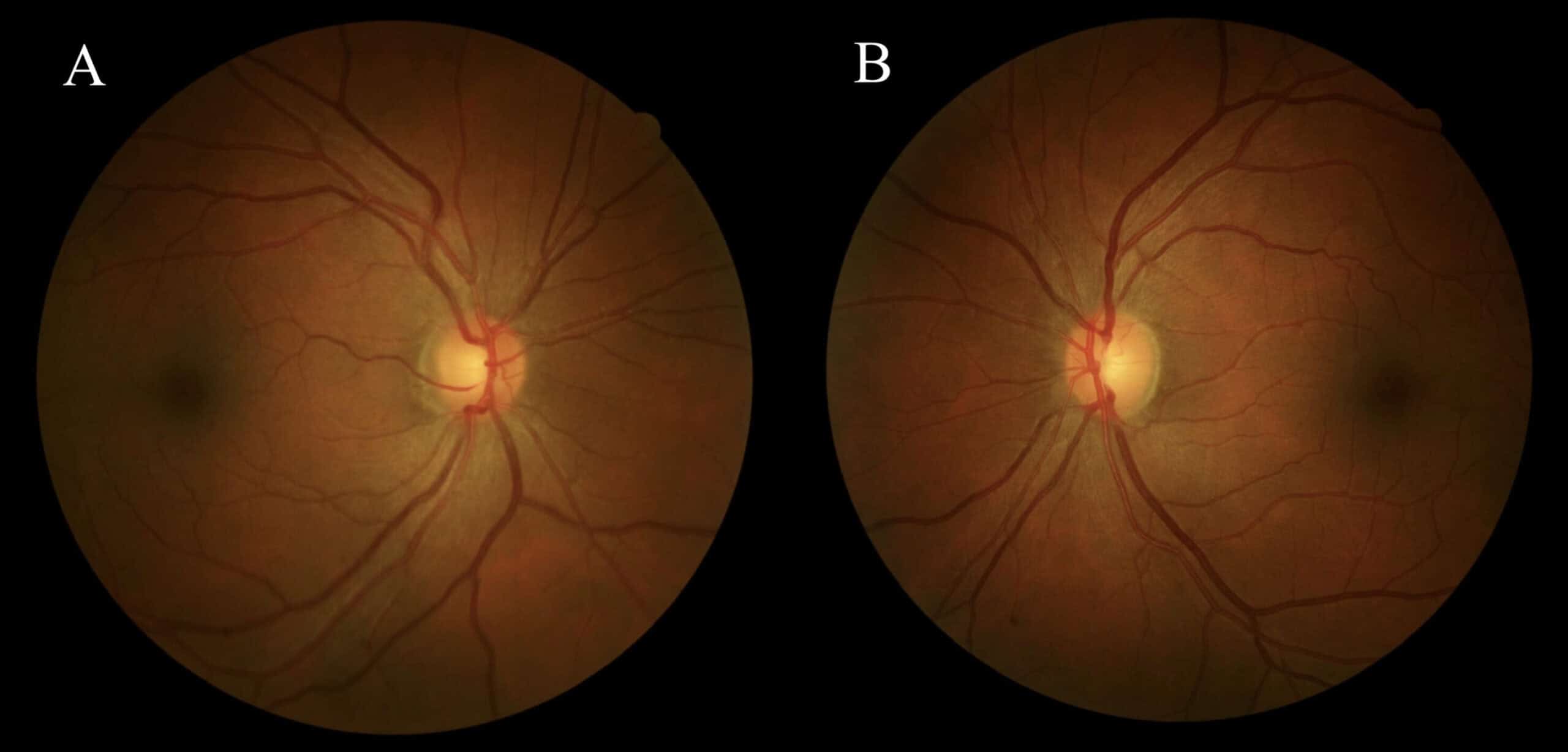
Figure 1: Fundus imaging of right eye (A) and left eye (B)
Annual OCT was acquired for monitoring low-risk glaucoma suspicion, demonstrating stability in the right eye and ~50 µm inferior temporal (IT) RNFL thickening of the left eye. Ganglion cell analysis was normal in both eyes.
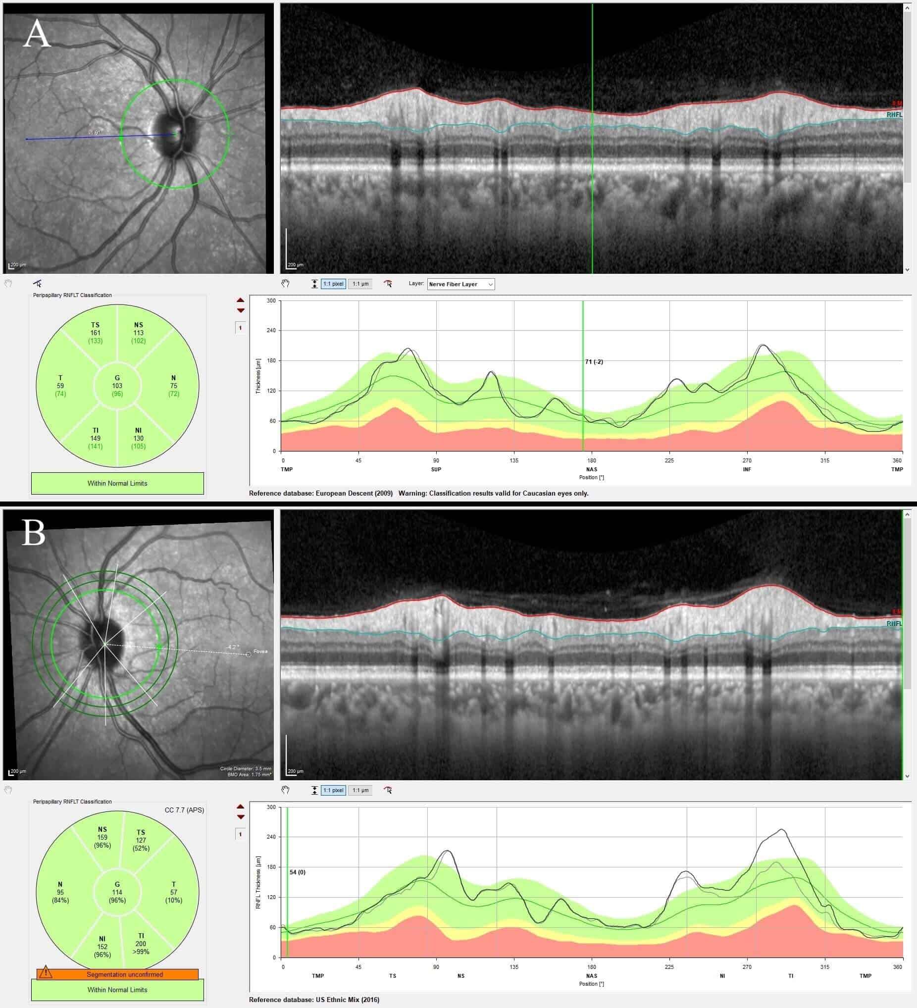
Figure 2: RNFL OCT of the right eye (A) and the left eye (B) with inferior temporal thickening in the left eye.
HVF testing was performed due to the sectoral RNFL thickening of the left eye, which revealed a corresponding, superior nasal visual field defect in the left eye.
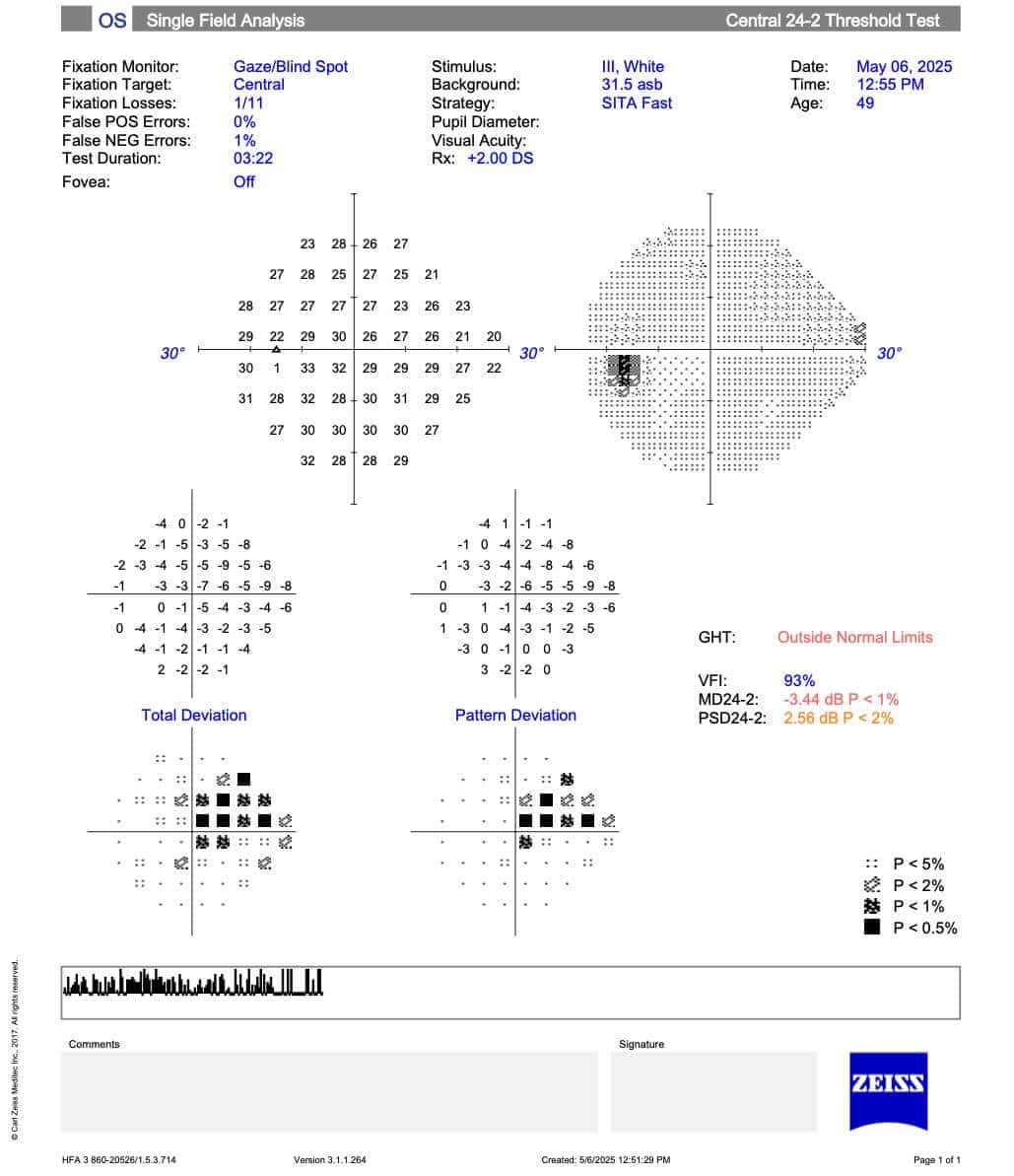
Figure 3: Visual Field of OS showing a dense superior nasal defect
In-office blood pressure was measured to be 119/71 mmHg. The patient denied any symptoms consistent with giant cell arteritis (GCA). The patient was diagnosed with suspected incipient subclinical sectoral non-arteritic anterior ischemic optic neuropathy of the left eye. Laboratory evaluation, including CBC, ESR, CRP, HbA1c, and infectious and inflammatory markers was normal. Given the patient’s history of bradycardia, the goal of minimizing nocturnal hypotension to reduce further ischemic risk and lifestyle modifications were reviewed. These included avoiding large meals and hot showers before bedtime, smoking cessation, maintaining a healthy diet, regular exercise, and medication compliance. After discussion with the patient’s primary care provider, aspirin 81 mg daily was initiated based on individualized cardiovascular risk assessment and low bleeding risk. A referral was also placed to re-establish care with the sleep clinic for reassessment of sleep apnea, given the patient’s prior borderline OSA diagnosis from eight years prior and its known association with NAION.
At the one-week follow-up visit, visual acuity, color vision, pupils, EOMs, OCT, and visual field were stable, with no worsening of the subclinical inferior temporal optic nerve edema in the left eye on RNFL OCT. IOPs measured 20 mmHg of the right eye and 19 mmHg of the left eye by GAT at 9:46AM. The patient remained asymptomatic and compliant with daily baby aspirin. At the one-month visit, the thickening of the RNFL OCT of the left eye had resolved to baseline with stable visual fields. IOPs measured 17 mmHg of the right eye and 19 mmHg of the left eye by GAT at 10:06AM. At the four-month visit, the left eye showed subsequent ~50 µm inferior temporal RNFL OCT thinning, mild improvement of the superior nasal defect on visual field testing, and questionable inferior temporal optic nerve pallor on clinical exam in the left eye. IOPs measured 20 mmHg of the right eye and 20 mmHg of the left eye by GAT at 11:40AM.
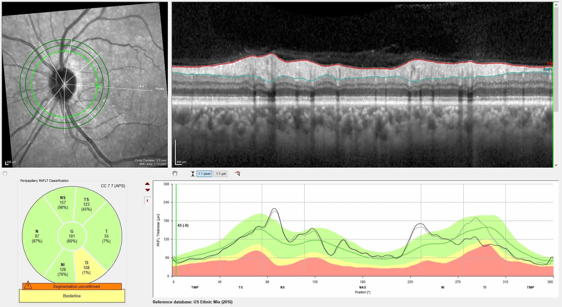
Figure 4: RNFL OCT OS four months after the event, showing inferior temporal thinning.
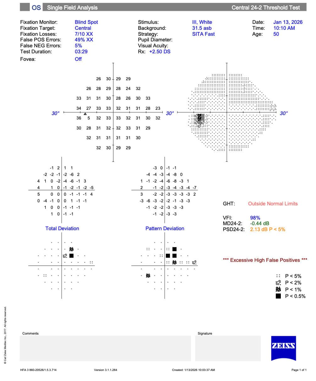
Figure 5: Visual Field of left eye four months after the event, showing an improved superior nasal defect (although somewhat unreliable)
At the seven-month visit, RNFL OCT of the left eye showed -7 µm RNFL thinning inferior temporal, and there remained questionable inferior temporal optic nerve pallor. Gonioscopy confirmed angle recession of 2 clock hours in the left eye. The IOPs measured 22 mmHg of each eye at 10:21 AM.
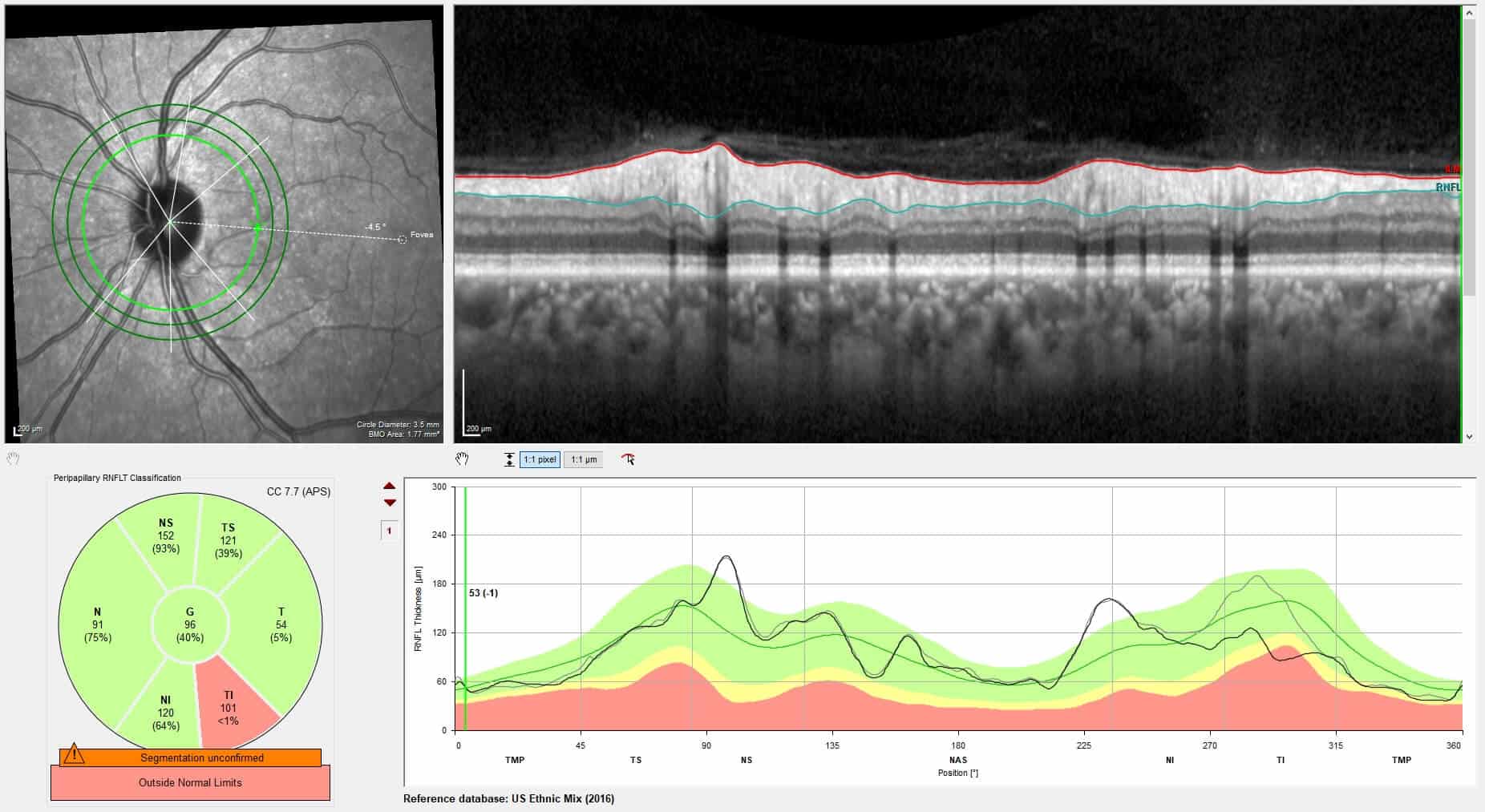
Figure 6: RNFL OCT of the left eye showing further inferior temporal thinning at seven months after initial presentation.
At the ten-month visit, RNFL OCT of the left eye remained stable without further thinning. The stability observed in the left eye without continued thinning supported a diagnosis of prior NAION over progressive glaucomatous neuropathy. IOPs were 20 mmHg in the right eye and 22 mmHg in the left eye at 1:12 PM, and moderate improvement of the superior nasal defect was shown on the visual field of the left eye. The nerves remained robust without pallor in the right eye, and questionable inferior temporal optic nerve pallor of the left eye.
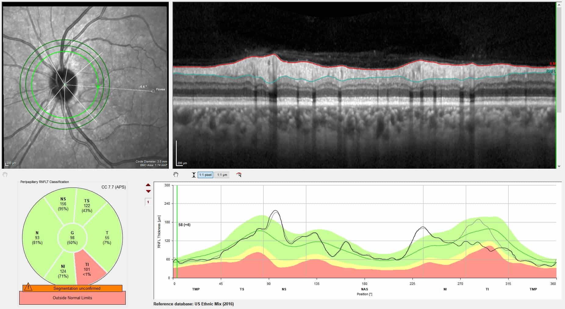
Figure 7: RNFL OCT of the left eye showing stable inferior temporal thinning between months seven and ten.
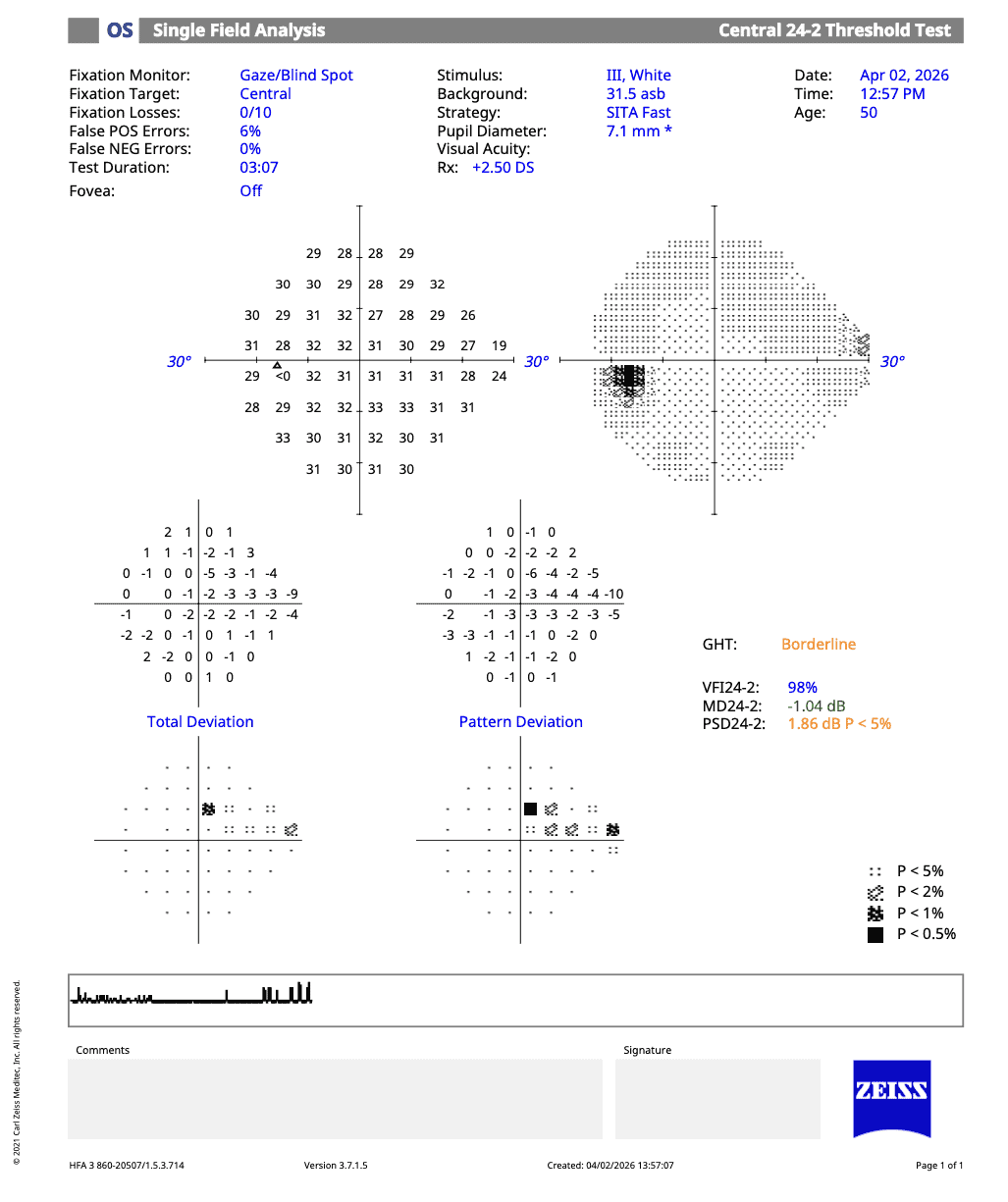
Figure 8: Visual Field of left eye ten months after the event showing a moderately improved superior nasal defect.
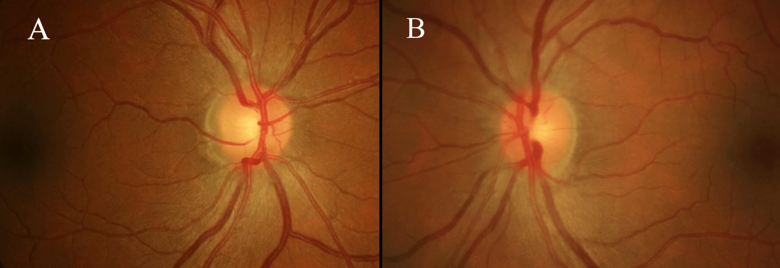
Figure 9: Fundus imaging of the right eye (A) and left eye (B) ten months after the event.
In the setting of average pachymetry readings (~540 µm in each eye), absence of worsening visual fields and OCTs, the patient’s diagnosis remained subclinical sectoral NAION of the left eye, and borderline ocular hypertension in both eyes with angle recession in the left eye. The patient is set to return for their comprehensive eye exam in five months.
Discussion
Differentiating NAION from glaucoma can be challenging, particularly when the ischemic event is mild, subclinical, sectoral, or evaluated outside of the acute phase. Both conditions result in retinal ganglion cell loss and visual field defects, but their pathophysiological mechanisms differ. NAION occurs due to acute hypoperfusion of the optic nerve, whereas glaucoma is associated with multifactorial chronic axonal degeneration.1,2
In this case, the presence of initial focal RNFL thickening on OCT in the left eye followed by resolution and subsequent thinning strongly supports an acute, ischemic process. In NAION, an acute ischemic event impairs mitochondrial function and axoplasmic transport in retinal ganglion cell axons. Mitochondrial dysfunction reduces ATP production required for ion transport, which disrupts axoplasmic flow leading to accumulation of cellular contents within the axon. This results in intracellular ionic imbalance, particularly sodium retention, driving water influx and resulting in cytotoxic axonal edema.6 Swelling occurs within the confined prelaminar and laminar optic nerve head in the scleral canal, and is hypothesized to compress the capillary circulation from the short posterior ciliary arteries and exacerbate ischemia, though the mechanism is not fully understood. Over time, apoptosis of retinal ganglion cells leads to permanent thinning of the RNFL and ganglion cell layer.6 Notably in this case, the optic disc appeared clinically unremarkable throughout the entire acute phase with no appreciable disc edema, peripapillary hemorrhages, or Paton’s folds, even as OCT revealed focal RNFL thickening. This case illustrates that sectoral NAION can be subclinical on fundus examination, underscoring the importance of ancillary imaging.
RNFL thickening in NAION is transient, typically occurring in the acute phase for a few weeks before resolving.4 Progressive RNFL thinning occurs for around four to six months as axonal loss evolves.4,6 Therefore, the observed RNFL thinning at seven months in this patient may still reflect the end of the natural course of post-ischemic structural loss rather than a glaucomatous process. Ongoing follow-up at ten months showed stability without further RNFL thinning, favoring a prior ischemic event over progressive glaucomatous change.
In contrast, typical chronic glaucomatous optic neuropathy does not demonstrate measurable RNFL thickening before thinning on OCT.4 Though the mechanism is not fully understood, glaucomatous optic neuropathy is driven by a multifactorial interplay of biomechanical, vascular, inflammatory, and neurodegenerative mechanisms that collectively result in progressive retinal ganglion cell loss.2,3,7,8 Damage to the lamina cribrosa, vascular dysregulation, IOP-related mechanical stress, translaminar pressure imbalance, neuroinflammation, and genetic susceptibility may also lead to glaucomatous changes, with chronic OCT thinning.2,3
As OCT is the current standard of care for monitoring both NAION and glaucoma, it plays a crucial role in distinguishing ischemic from glaucomatous damage. Had the patient presented only after resolution of RNFL thickening of the left eye, the residual thinning and field defect could have been misinterpreted as glaucomatous damage, potentially prompting unnecessary IOP-lowering therapy. Longitudinal imaging identified transient RNFL edema followed by structural loss, consistent with NAION.4,6
Visual field findings differ between the two conditions. NAION-related defects may show partial improvement or fluctuation following resolution of acute disc edema from recovery of reversibly injured axons. In contrast, glaucomatous visual field defects are typically progressive and irreversible.4,5 The moderate improvement observed in this patient’s visual field defect in the left eye following resolution of RNFL edema supports an ischemic etiology.
This patient’s systemic and ocular risk profile further complicated diagnostic interpretation. Borderline OSA and bradycardia may contribute to nocturnal optic nerve hypoperfusion, increasing susceptibility to ischemic injury.9-10 In one study, NAION patients are approximately 4.9 times more likely to have obstructive sleep apnea compared to controls, and patients with untreated sleep apnea have a 16% increased risk of developing NAION.1 Fellow-eye involvement occurs in 15–24% of cases over five years, underscoring the need for systemic risk factor modification.1 Evidence linking OSA and glaucoma is mixed, with some studies showing a modest relative increase of ~40% risk, while others show no significant association.11 Given the patient’s relatively low glaucomatous risk profile, the absolute increase in risk from OSA is likely small.
African American race and prior ocular trauma with angle recession are established risk factors for glaucoma.12 Angle recession can damage the trabecular meshwork and increase the risk of delayed IOP elevation.13 While only about 5% of patients with isolated angle recession develop glaucoma, among those with unilateral angle-recession glaucoma, approximately 50% develop primary open-angle glaucoma in the fellow eye over time.14 Although the relative risk is increased, the baseline risk remains low. Given the coexistence of risk factors for both NAION and glaucoma, and the potential for overlapping ischemic and glaucomatous mechanisms, clinicians should interpret imaging longitudinally and carefully. Pre-existing damage from one condition may confound assessment of progression in the other.
Management of NAION focuses on systemic risk factor modification, as no therapy has been shown to reverse optic nerve damage.1 Low-dose aspirin has been studied as a strategy to reduce the risk of fellow eye involvement, although evidence remains inconclusive.15 In clinical practice, the decision to initiate aspirin is often individualized and typically considered when the patient has many vascular risk factors and a low risk of bleeding complications. In this case, co-management with the patient’s primary care provider led to the decision that starting aspirin 81 mg daily was reasonable given its potential protective effect on the fellow eye. In contrast, glaucoma management is directed toward lowering intraocular pressure to prevent progressive axonal loss. NAION and glaucoma are not mutually exclusive conditions and may coexist within the same eye, making pre-existing damage from one condition harder to detect changes from the other.
This case highlights the importance of integrating imaging findings within the patient’s full clinical context, including age, optic nerve anatomy, systemic vascular risk factors, ocular history, and disease timeline, rather than relying solely on isolated OCT thinning or visual field defects. However, in very mild or sectoral events, clinical evidence may be difficult to appreciate. In patients with overlapping risk factors, clinicians should avoid a premature diagnosis based on a single visit or a structural parameter alone. An individualized approach can help avoid unnecessary treatment in young patients with uncertain glaucomatous change while still allowing for appropriate monitoring over time. Eye care providers must recognize these imaging patterns to guide appropriate evaluation, systemic risk assessment, and longitudinal monitoring in patients with suspected glaucomatous or ischemic optic neuropathy.
References
- Hayreh SS. Ischemic optic neuropathies—where are we now? Graefes Arch Clin Exp Ophthalmol. 2013;251(8):1873–1884.
- Jonas JB, Aung T, Bourne RR, Bron AM, Ritch R, Panda-Jonas S. Glaucoma. Lancet. 2017;390(10108):2183–2193.
- Weinreb RN, Khaw PT. Primary open-angle glaucoma. Lancet. 2004;363(9422):1711–1720.
- Contreras I, Rebolleda G, Noval S, Muñoz-Negrete FJ. Optic disc evaluation by OCT in NAION. Invest Ophthalmol Vis Sci. 2007;48(9):4087–4092.
- Hood DC, Kardon RH. Framework for comparing structural and functional glaucomatous damage. Prog Retin Eye Res. 2007;26(6):688–710.
- Arnold AC. Pathogenesis of nonarteritic anterior ischemic optic neuropathy. Surv Ophthalmol. 2018;63(1):1–15.
- Fortune B, Cull GA, Reynaud J, Wang L. Relating retinal ganglion cell loss to RNFL thickness in glaucoma. Prog Retin Eye Res. 2017;58:1–18.
- Leung CKS, Ye C, Weinreb RN, et al. RNFL imaging with SD-OCT: patterns of progression in glaucoma. Ophthalmology. 2018;125(5):701–710.
- Palombi K, Renard E, Levy P, et al. NAION and obstructive sleep apnea association. Br J Ophthalmol. 2006;90(7):879–882.
- Archer E, Pepin S. Obstructive sleep apnea and NAION. J Clin Sleep Med. 2018;9:613–618.
- Cheong AJY, Wang SKX, Woon CY, et al. Obstructive sleep apnea and glaucoma: a systematic review and meta-analysis. Eye (Lond). 2023;37(15):3065–3083.
- Tielsch JM, Sommer A, Katz J, Royall RM, Quigley HA, Javitt J. Racial variations in POAG prevalence. JAMA. 1991;266(3):369–374.
- Salmon JF, Mermoud A, Ivey A, Swanevelder SA, Hoffman M. Detection of post-traumatic angle recession. Ophthalmology. 1994;101(10):1846–1850.
- Tesluk GC, Spaeth GL. Primary open-angle glaucoma occurrence in the fellow eye. Ophthalmology. 1985;92(7):904–911.
- Stiebel-Kalish H, Hasanreisoglu M, Leibovici L. Aspirin following NAION: systematic review and meta-analysis. Eye. 2010;24(6):1015–1023.

Dr. Emily Coan graduated from the Southern California College of Optometry in 2025 and completed a residency in Ocular Disease at the VA Boston Healthcare System, Jamaica Plain Campus. She recently joined the Neuro-Ophthalmology and Primary Care Optometry Services at Massachusetts Eye and Ear.

Dr. Jordan graduated from The New England College of Optometry in 2017 with a combined OD/MS degree. She then completed an ocular disease/primary care residency at the Boston VA, Jamaica Plain campus from 2017-2018, where she is now an attending optometrist and contact lens clinic lead. Her hobbies include reading, spending time with her family, and going to the beach.