

Kearns-Sayre Syndrome with a Finding of Unique Subretinal Hyperreflective Deposits: A Case Report
 Save as PDF
Conflict of interest: The author(s) declared that this work was conducted in the absence of any commercial or financial relationships that could be construed as a potential conflict of interest.
Abstract
Background: Kearns-Sayre syndrome is a rare mitochondrial DNA deletion disorder characterized by the body’s multisystem involvement, including the eyes. This report follows the disease progression of a patient with Kearns-Sayre syndrome (KSS). It documents a novel presentation of unique multiple subretinal hyperreflective deposits, expanding the phenotypic spectrum of ocular manifestations associated with this syndrome.
Case Report: A 16-year-old Asian male with Kearns-Sayre syndrome was regularly evaluated to monitor the progression of his ocular symptoms, which included ptosis, chronic progressive ophthalmoplegia, and pigmentary retinopathy. The patient’s clinical course, imaging studies, and management plans are detailed. A thorough ocular exam, including retinal imaging, confirmed the presence of unique multiple subretinal hyperreflective deposits that are distinct from the characteristic retinopathy seen in Kearns-Sayre syndrome.
Conclusion: The identification of multiple subretinal hyperreflective deposits in this patient with Kearns-Sayre syndrome highlights a rarely reported manifestation of the disease. Our findings may suggest that subretinal hyperreflective deposits may be an under-recognized feature. This case demonstrates the importance of vigilant ocular examination and broadens understanding of ocular complications associated with Kearns-Sayre syndrome. Further research into the mechanisms driving this manifestation could yield new insights into Kearns-Sayre syndrome’s mitochondrial pathology and its impact on the retina.
Keywords: Kearns-Sayre syndrome, subretinal deposits, mitochondrial disorders, pigmentary retinopathy, progressive external ophthalmoplegia
Introduction
Kearns-Sayre syndrome (KSS) is a rare and severe genetic disorder, characterized by a spectrum of signs and symptoms resulting from mutations in relation to mitochondrial DNA.1 Kearns-Sayre syndrome can result from large-scale deletions of mitochondrial DNA or from mutations in the nuclear DNA that regulate mitochondrial DNA and code for mitochondrial proteins.1,2 Mitochondria are critical for cellular energy production, and their malfunction causes detrimental consequences, especially in high-energy-demanding tissues such as the nervous system and muscles, including the retina, ocular muscles, and the heart.3,4 Kearns-Sayre Syndrome was first clinically defined in 1958 by Thomas P. Kearns and George Pomeroy Sayre, who identified its hallmark triad: progressive external ophthalmoplegia, pigmentary retinopathy, and onset before the age of 20.5 This disease played an important part in understanding the correlation between mitochondrial DNA deletions and their clinical phenotypes.4,6
Case Report
Initial Visit
A 10-year-old Asian male presented to the eye clinic with his mother. He was a new patient, and referred by an outside optometrist for suspected retinitis pigmentosa. Translation services were used to obtain case history from the patient’s mother, who spoke Mandarin, but still, communication was limited. The patient complained of blurry vision, worsening over the last six months, which made it difficult for him to read fine print at school. The complaint of blurred vision was in both eyes, at distance and near and was constant. Upon further questioning, the patient also reported poor night vision (nyctalopia), light sensitivity (photophobia), and drooping of both upper eyelids (ptosis), which the mother indicated that the worsening had been noticed months ago. When asked, he denied bumping into objects and having tunnel vision.
Pertinent medical history included gastrointestinal issues that caused the patient not to eat well, having a short stature, falling often due to muscle weakness, and not performing well academically in school. The patient had a normal birth, no additional digits, normal face shape, and normal hearing. The patient’s entering uncorrected visual acuity was 20/50 in the right eye and 20/100 in the left eye. He was unable to perform a pinhole acuity test due to comprehension. The patient scored 0 out of 6 on Hardy-Rand-Rittler (HRR) color vision testing in each eye. Using the Mars contrast sensitivity test, he had moderate loss of contrast sensitivity in the right eye and severe loss in the left eye. Pupils were equally round, reactive to light, and without afferent pupillary defects. The confrontation visual field test result was full to finger count in all quadrants for the right eye, and questionably restricted in the superonasal quadrant in the left eye, with poor fixation. Extraocular muscle movements were full. After manifest refraction, it was found that the patient had simple myopic astigmatism in each eye, and his best corrected visual acuity was 20/40 in the right eye and 20/60 in the left eye. The patient had bilateral ptosis with a 7 mm palpebral aperture in each eye.
Anterior segment findings were otherwise unremarkable. Intraocular pressure was normotensive in both eyes.
The lens of each eye was clear. Posterior segment evaluation revealed normal vitreous, unremarkable optic nerve heads with small cupping, and clear maculae in both eyes. Retinal arteries appeared to be mildly attenuated, while the veins were unremarkable in both eyes. In the mid-periphery and peripheral retina, pigmentary changes were noted bilaterally in all quadrants. Optical coherence tomography (OCT) of the macula revealed thinning of the outer retinal layers, with a faint external limiting membrane and ellipsoid zone and a jagged interdigitation zone in both eyes. In addition, there was widespread thinning and irregularity of the RPE. There was no intraretinal fluid, subretinal fluid, or choroidal neovascular membrane. OCT also demonstrated a normal retinal nerve fiber layer thickness in all quadrants of the right eye; the patient was unable to fixate to obtain an image for the left eye.
At this visit, the patient was diagnosed with pigmentary retinopathy with suspected rod-cone dystrophy vs. cone-rod dystrophy. While both subtypes of pigmentary retinopathies lead to eventual atrophy of both cone and rod photoreceptors, the patient was only in his second decade of life. The patient was scheduled for an electroretinogram to determine the diagnosis. He was also given a spectacle prescription for full-time wear and was referred to the low vision clinic for evaluation.
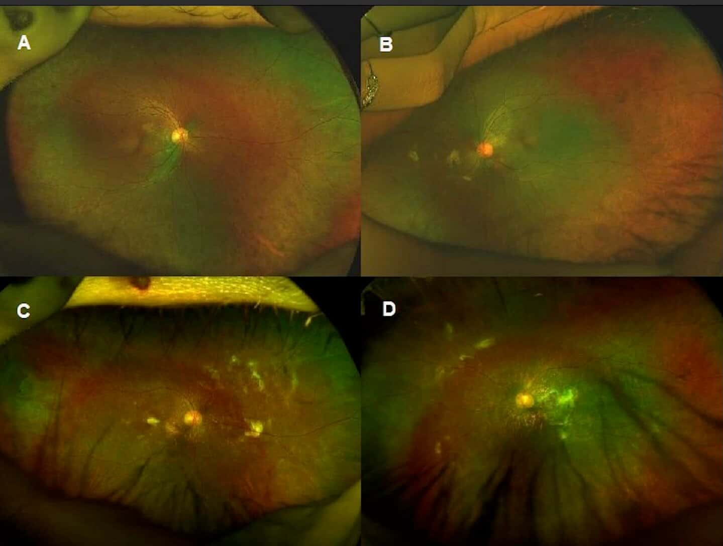
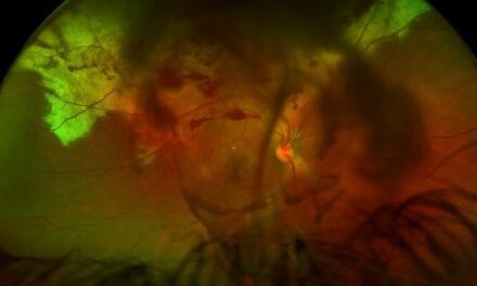
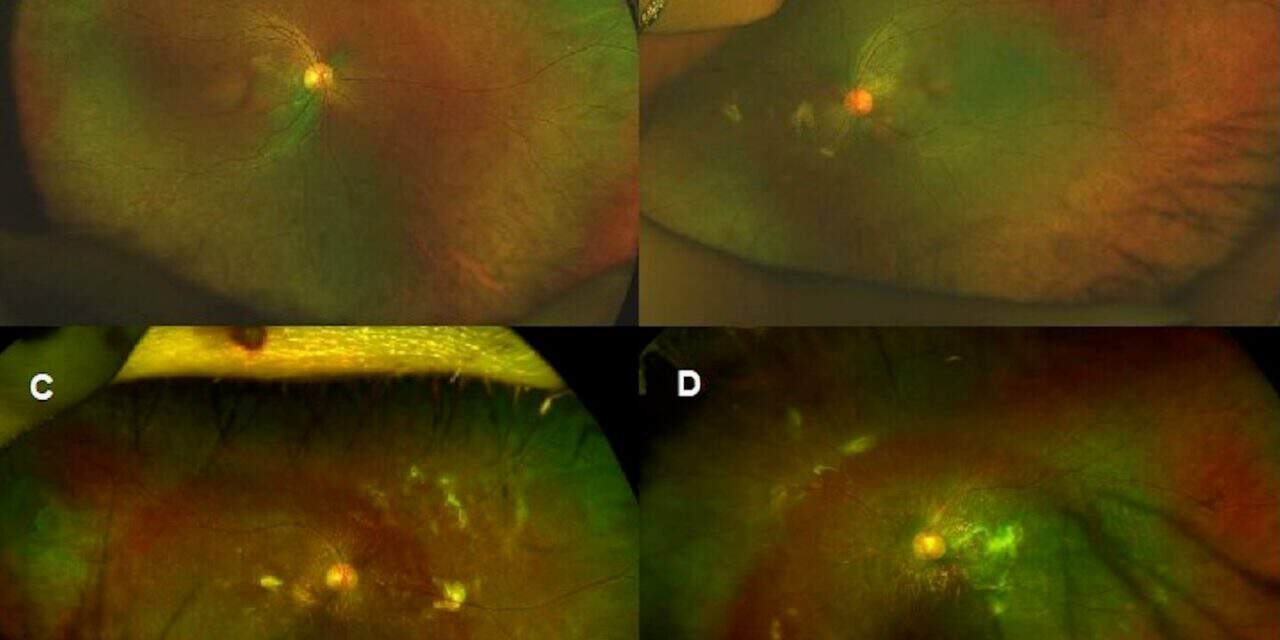
Figure 1. Color fundus photos, A-B taken at initial visit, C-D 6 years after the initial visit. A: Right eye, attenuated arterioles with pigmentary retinopathy. B: Left eye, attenuated arterioles with pigmentary retinopathy. C: Demonstrating additional multiple subretinal hyperreflective lesions in the right eye 6 years after the initial visit. D: Demonstrating additional multiple subretinal hyperreflective lesions in the left eye.
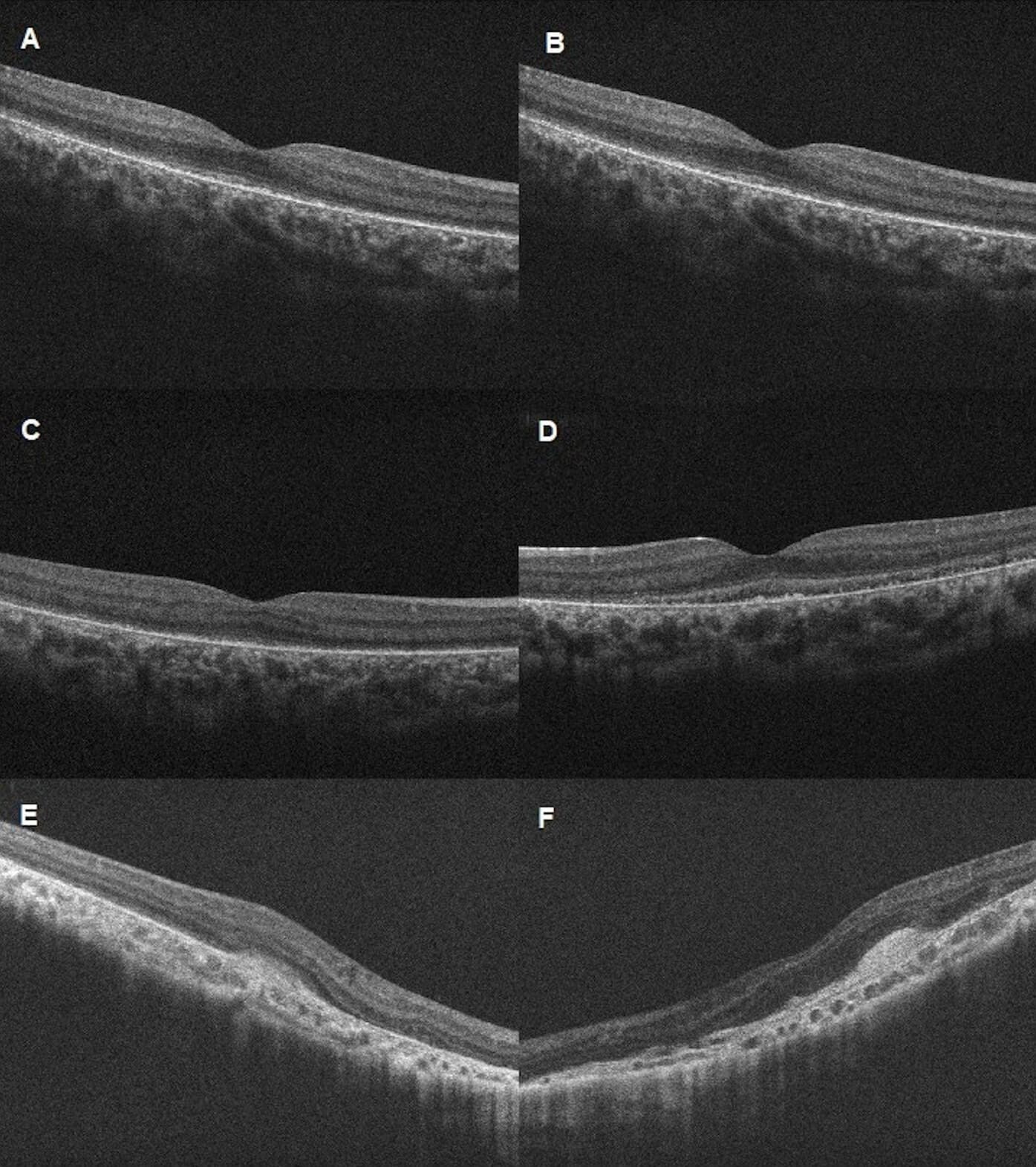
Figure 2. HD-Raster optical coherence tomography through the fovea. A-B taken at initial visit, C-D 1 years after the initial visit, E-F 6 years after the initial visit. A: Right eye, faint outer limiting membrane and photoreceptor line, and jagged interdigitation zone. B: Left eye, faint outer limiting membrane and photoreceptor line, and jagged interdigitation zone. C: Right eye, absence of the photoreceptor integrity line and outer retinal thinning. D: Left eye, absence of the photoreceptor integrity line and outer retinal thinning. E: Right eye, subretinal hyperreflective lesions. F: Left eye, subretinal hyperreflective lesions.
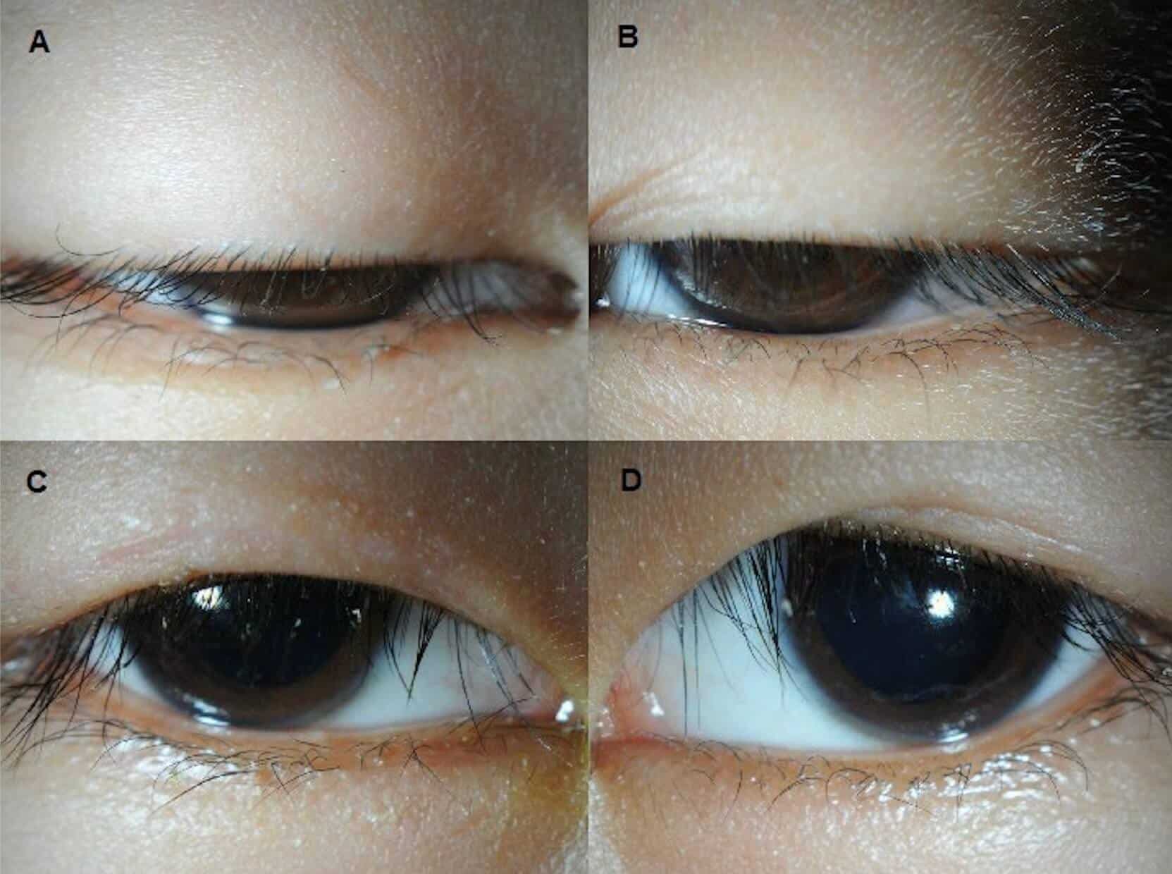
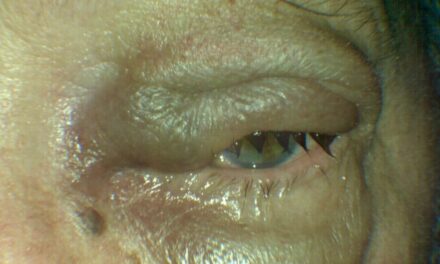
Figure 3. Anterior segment photos taken with the patient looking straight ahead OU. A-B taken at initial visit, C-D after surgical ptosis repair.
A: Right eye with severe ptosis. B: Left eye with severe ptosis. C: Right eye. D: Left eye.
Follow-up, 10 months after initial visit
At this visit, ten months later, the patient presented with his mother and showed a worsening ptosis in both eyes and a new diagnosis of celiac disease. The patient had a stable best-corrected visual acuity of 20/40 and 20/60 in the right and left eye, respectively. His margin to reflex distance 1 (MRD1) was 0.5 mm in the right eye and negative in the left eye.
Levator function was measured and was poor in each eye. All other external findings were stable. The patient also had mild papillae on both the upper and lower palpebral conjunctivae. All other anterior segment findings remained stable during his initial visit. The patient’s intraocular pressures were normotensive. Dilated posterior segment findings were stable from the initial visit. OCT of the macula showed progressive loss of the ellipsoid zone and interdigitation zone compared to the previous scan. The left eye also demonstrated a shallow, diffuse, neurosensory detachment. This finding of increased distance between the ellipsoid zone and the RPE is characteristic of a common phenotype of mitochondrial retinopathies. Thickness mapping showed diffuse macular thinning in the right eye and relative stability in the left eye. The patient also performed a visual field test using Octopus 900 Perimeter. The G pattern tendency-oriented perimetry (G-TOP) report showed a generalized deep depression and restriction of all quadrants with inferonasal sparing in the right eye and a generalized depression of all quadrants that were worse in the inferonasal region of the left eye. Last, electroretinogram testing showed extinguished rod, rod-cone, and cone responses, with minimal flicker response remaining in both eyes.
At this visit, the patient was diagnosed with significant ptosis, allergic conjunctivitis, and generalized pigmentary retinopathy of both eyes. He was prescribed ketotifen 0.025% ophthalmic solution for the allergic conjunctivitis. Ptosis surgery was discussed, and the patient was referred to an oculoplastic surgeon for consultation. Given the patient’s medical history, symptoms, and clinical findings, Kearns-Sayre syndrome was suspected, and genetic testing with the patient’s pediatrician was recommended. The patient was advised to return in six months for follow-up in our retina clinic, and to continue care in the low vision clinic.
Follow-up, 4 years after initial visit
The patient had a series of follow-up visits in the anterior segment, low vision, and retina clinics. The patient had surgical repair of the ptosis with a bilateral frontalis suspension procedure two years prior. Genetic testing had been performed which confirmed the diagnosis of Kearns-Sayre syndrome. A formal letter was sent to the eye clinic stating the diagnosis based on genetic testing, and the mother confirmed verbally that the genetic testing results were positive for Kearns-Sayre syndrome. However, despite multiple attempts to obtain the detailed report of the genetic testing, we were unsuccessful.
He reported decreasing vision of both eyes and difficulty seeing things in the classroom. Although he did not report any falls or injuries, he did report bumping into objects, especially at night. At this visit, the best corrected visual acuity was stable at 20/50 in the right eye and 20/60 in the left eye. Extraocular movements were now restricted to superior and inferior gazes in each eye. MRD-1 was improved to 3 mm in the right eye and 2 mm in the left eye. Anterior segment examination revealed mild scurf with inferior punctate staining in both eyes, incomplete blink OU and incomplete nasal eyelid closure of the left eye. All other external and anterior segment findings were stable. Intraocular pressures were normotensive. Dilated fundus findings remained stable compared to the previous, including the optical coherence tomography results for both eyes.
The patient had developed early signs of ophthalmoplegia in both eyes and blepharitis with superficial punctate keratitis in both eyes. The ptosis repair surgery in both eyes led to a secondary incomplete lid closure in the left eye. Treatments for dry eyes, such as lid hygiene techniques, artificial tears, and erythromycin ointment, were prescribed for the patient. Regular follow-ups in the anterior segment, low vision, and retina clinics were recommended.
Follow-up, 6 years after initial visit
After being lost to follow-up for two years, the now 16-year-old patient returned to the clinic reporting vision loss over the last few years. The patient’s mother stated that the patient was now also regularly followed by a cardiologist and an endocrinologist every six months. The patient’s best corrected visual acuity worsened to 20/120 for the right eye, and 1/600 for the left eye. Confrontation visual fields were restricted in all quadrants, and extraocular muscles were restricted in all gazes in both eyes, rendering the patient’s eyes fixed in primary gaze. The patient had relatively stable MRD values in both eyes. Anterior segment findings included matted lashes, inferior corneal pannus in both eyes, mild inferior punctate keratitis in the right eye, and significant inferior punctate keratitis in the left eye. He also had a heaped epithelium and a 1 mm corneal infiltrate off the visual axis in the left eye. All other external and anterior segment findings were stable compared to the previous. The patient’s intraocular pressures were stable.
During dilated fundus evaluation, it was found that the patient had developed multiple subretinal deposits in the macula, mid-periphery, and periphery. Optical coherence tomography was then performed to investigate the presence of subretinal deposits in the macula of each eye. At this visit, the patient was diagnosed with marginal keratitis in the left eye, chronic progressive ophthalmoplegia, and pigmentary retinopathy with multiple subretinal hyperreflective deposits in both eyes. Tobramycin 0.3% and dexamethasone 0.1% ophthalmic suspension QID was prescribed in the left eye for the marginal keratitis, and cyclosporine 0.09% ophthalmic emulsion BID in both eyes for the bilateral keratitis. He was advised to restart preservative-free artificial tears, lid hygiene, and lid tape OU at night. The patient and his mother were educated that there was no current treatment for the subretinal deposits, and it was advised to monitor closely with an OCT of the macula.
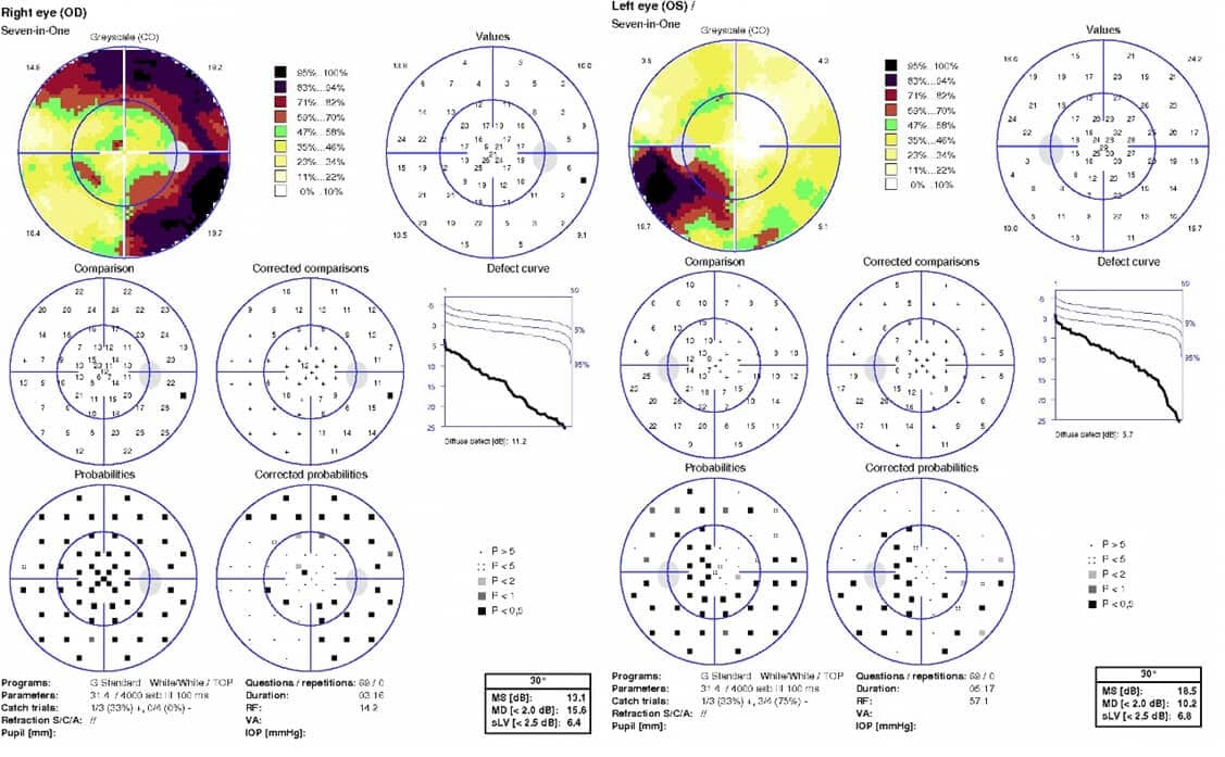
Figure 4. G-TOP reports one year after first visit. Results show superior, temporal, and inferior constriction in the right eye and generalized constriction with a worse inferotemporal quadrant in the left eye.
Discussion
Before specifically discussing Kearns-Sayre syndrome and its ocular effects, it is important to recognize the wide array of systemic manifestations caused by mitochondrial dysfunction that identify this disorder. Because mitochondria play an important role in providing energy to muscle cells in the heart, the system’s irregular heartbeat control can cause heart block.5 Various other muscles are affected as well, including muscles of the digestive system, respiratory muscles, and skeletal muscles.1 Dysphagia and gastroparesis can cause poor feeding and nutrition due to the difficulty of food consumption.7 When the respiratory muscles are involved, respiratory insufficiency can occur, especially during sleep. Skeletal muscle involvement causes general mobility problems. In addition, neuropathies such as ataxia and peripheral neuropathy can further affect patient coordination.1 Sensorineural hearing loss can occur at mild to profound levels, and patients often require hearing aids.1,4 The endocrine system is also affected by Kearns-Sayre syndrome. Hypoparathyroidism has been documented and can lead to hypocalcemia. Diabetes mellitus can also occur, as well as growth hormone deficiencies, causing short stature and delayed puberty in some cases.8
Ocular involvement is particularly significant in Kearns-Sayre syndrome and is known to be bilateral and symmetrical. Progressive external ophthalmoplegia is a hallmark feature of Kearns-Sayre syndrome, characterized by worsening ptosis and progressive weakness or paralysis of the extraocular muscles, eventually leading to immobile eyes.1,5 Ptosis surgery caused the secondary incomplete closure of the left eyelid, resulting in exposure keratopathy, which indicated the need for ocular surface treatments.
Another hallmark ocular sign is pigmentary retinopathy.1,5 While differentiated by the genetic origin and the pathophysiology, the signs and symptoms are very similar to those of retinitis pigmentosa. Patients will have a varying degree of gradual worsening of nyctalopia, peripheral vision loss, and often later, central vision loss.1,9 For this patient, exam findings indicated evidence of both cone and rod photoreceptor atrophy, warranting further investigation with electroretinography. Because the electroretinogram showed extinguished photoreceptor responses across all parameters, the patient was diagnosed with generalized pigmentary retinopathy. Correct evaluation of these ocular manifestations not only aids in the identification of Kearns-Sayre syndrome, but also plays a crucial role in patient care given the potential for significant impact on the patient’s quality of life due to the many systemic comorbidities.
In this case report, a 16-year-old Asian male with Kearns-Sayre syndrome displayed the expected course of ocular and systemic signs and symptoms, with the addition of ocular manifestations of unusual subretinal deposits. While the pigmentary retinopathy did significantly affect the patient’s vision, his reduced best-corrected visual acuity after two years of being lost to follow-up was mainly due to the subretinal deposits in the fovea in both eyes. Although subretinal deposits are characteristic of inherited retinal degenerations, the ones that appear in this case have a unique hyperreflective appearance. Although the nature of these deposits has not yet been identified, the white, wispy, non-uniform appearance of these deposits has not been reported in Kearns-Sayre. To our knowledge, there has been one other reported patient with Kearns-Sayre syndrome who developed subretinal complications. There was a reported case of subretinal fibrosis in both eyes and a reported case of persistent subretinal fluid in both eyes.10,11
Both of these patients followed the expected course of pigmentary retinopathy, external ophthalmoplegia, and heart block under the age of 20, similar to the patient in this case report. Whether the addition of subretinal change was a coincidental finding or a unique advanced manifestation of the syndrome in these patients remains a topic for discussion. The patient in this case report had no evidence of retinal detachment or choroidal neovascularization, which rules out common causes of fibrotic tissue development. Pigmentary retinopathy is inherently a low-grade chronic inflammatory process of cell atrophy, so it is speculated that his presentation of unique subretinal hyperreflective deposits are from an inflammatory origin.12 Supporting this hypothesis is the findings of a histopathologic study of a KSS patient which demonstrated that the deposits may consist of a mixture of RPE-photoreceptor degenerated cellular elements rather than a simple drusen-like deposit. In addition, the surviving photoreceptor–RPE islands may contain large pigment-laden macrophages in the subretinal space together with photoreceptor outer-segment debris.13 The white color seen in these lesions may therefore represent an active chronic immune response. Macrophages inefficiently clear debris due to ATP depletion, as seen in KSS. The remaining cellular inflammatory aggregates scatter light strongly, resulting in a white appearance.
Despite differences in pathophysiology, it is important to keep in mind that subretinal fibrosis is a novel phenotype in other conditions that cause atrophy of the retinal pigmented layer and choroid, such as Goldman-Favre syndrome, choroideremia, or Stargardt disease.14,15,16 Despite well-documented associations between Kearns-Sayre syndrome and ocular abnormalities, the spectrum of ocular manifestations continues to expand, highlighting the variable nature of mitochondrial diseases. The documentation of novel ocular findings in Kearns-Sayre syndrome patients contribute to the clinical understanding of the syndrome and challenge clinicians and researchers to adapt their diagnostic and therapeutic approaches.
Conclusion
Kearns-Sayre syndrome is a rare mitochondrial DNA deletion disorder characterized by the body’s multisystem involvement, including the eyes. Careful ocular examination and the use of advanced imaging technology are crucial for managing these patients. This report documents a presentation of unique subretinal hyperreflective deposits, expanding the phenotypic spectrum of ocular manifestations associated with this syndrome. Additional research is essential to investigate and develop effective treatments for macular subretinal deposits that can lead to visual impairment. This case report aims to enhance the understanding of ocular complications related to Kearns-Sayre Syndrome and highlights the importance of interprofessional collaboration.
The authors would like to acknowledge Shaoheng Chen, OD, MS, for his contribution regarding the nature of the subretinal deposition in KSS.
References
- Goldstein A, Falk MJ. Single Large-Scale Mitochondrial DNA Deletion Syndromes. In: Adam MP, Feldman J, Mirzaa GM, Pagon RA, et al., eds. GeneReviews®. Seattle (WA): University of Washington, Seattle; 2003 [updated 2023 Sep 28].
- Broomfield A, Sweeney MG, Woodward CE, Fratter C, Morris AM, Leonard JV, et al. Paediatric single mitochondrial DNA deletion disorders: an overlapping spectrum of disease. J Inherit Metab Dis. 2015;38:445-57.
- Reynolds E, Byrne M, Ganetzky R, Parikh S. Pediatric single large-scale mtDNA deletion syndromes: The power of patient reported outcomes. Mol Genet Metab. 2021;134:301-8.
- Mancuso M, Orsucci D, Angelini C, Bertini E, Carelli V, Comi GP, et al. Redefining phenotypes associated with mitochondrial DNA single deletion. J Neurol. 2015;262:1301-9.
- Kearns TP, Sayre GP. Retinitis Pigmentosa, External Ophthalmoplegia, and Complete Heart Block: Unusual Syndrome with Histologic Study in One of Two Cases. AMA Arch Ophthalmol. Aug 1958;60(2):280-9.
- Zeviani M, Moraes CT, DiMauro S, Nakase H, Bonilla E, Schon EA, et al. Deletions of mitochondrial DNA in Kearns-Sayre syndrome. Neurology. 1988;38(9):1339-1346.
- Finsterer J, Frank M. Gastrointestinal manifestations of mitochondrial disorders: a systematic review. Therap Adv Gastroenterol. 2017;10(1):142-154.
- Al-Gadi IS, Haas RH, Falk MJ, Goldstein A, McCormack SE. Endocrine disorders in primary mitochondrial disease. J Endocr Soc. 2018;2:361-73.
- O’Neal TB, Luther EE. Retinitis Pigmentosa. [Updated 2024 Feb 12]. In: StatPearls [Internet]. Treasure Island (FL): StatPearls Publishing; 2024 Jan.
- Kozak I, Oystreck DT, Abu-Amero KK, Nowilaty SR, Alkhalidi H, Elkhamary SM, et al. New Observations Regarding the Retinopathy of Genetically Confirmed Kearns-Sayre Syndrome. Retina Cases Brief Rep. 2018;12(4):349-358.
- Paulus YM, Wenick AS. Development of Chronic Subretinal Fluid in Kearns-Sayre Syndrome. Retin Cases Brief Rep. 2016;10(3):236-238.
- Kaur G, Singh NK. Inflammation and retinal degenerative diseases. Neural Regen Res. 2023;18(3):513-518.
- McKechnie NM, King M, Lee WR. Retinal pathology in the Kearns-Sayre syndrome. Br J Ophthalmol. 1985 Jan;69(1):63-75.
- Alsalamah AK, Khan AO, Bakar AA, Schatz P, Nowilaty SR. Recognizable Patterns of Submacular Fibrosis in Enhanced S-Cone Syndrome. Ophthalmol Retina. 2021;5(9):918-927.
- Endo K, Yuzawa M, Ohba N. Choroideremia associated with the subretinal neovascular membrane. Acta Ophthalmol Scand. 2000;78(4):483-486.
- Rossi S, Testa F, Attanasio M, Orrico A, et al. Subretinal Fibrosis in Stargardt’s Disease with Fundus Flavimaculatus and ABCA4 Gene Mutation. Case Rep Ophthalmol. 2012;3(3):410-417.

Dr. Canellos is a full-time faculty member of SUNY State College of Optometry. She is an Associate Clinical Professor, Instructor of Record of the Fourth Year Program and the Director of the University Eye Center’s Referral Service. She supervises interns and residents in the Anterior Segment/Cataract clinics of the Advanced Care Service.

Dr. Bass is a Distinguished Teaching Professor and a 45-year member of the faculty at the SUNY College of Optometry. She is an attending in the Retina and Electrodiagnostic Clinic and Residency Supervisor of the SUNY Residency in Ocular Disease.

Dr. Inwhan Lee is an optometrist at Kaiser Permenente Woodland Hills. Dr. Lee is a graduate of Western University College of Optometry and has finished residency program in ocular disease at SUNY College of Optometry.