

Problematic Proptosis

 Save as PDF
Conflict of interest: The author(s) declared that this work was conducted in the absence of any commercial or financial relationships that could be construed as a potential conflict of interest.
Welcome to the “Neuro Nuggets” column within the Journal of Medical Optometry (JoMO)! This column aims to make neuro-ophthalmic disease more approachable by blending real-world clinical cases with evidence-based medicine. The patient’s case in this edition’s column demonstrates the importance of recognizing proptosis as a heralding feature of orbital (and often systemic) disease. Enjoy!
Case Presentation
An 81-year-old male was admitted to the hospital for weakness in his extremities and binocular diplopia. His past medical history included prior prostate cancer s/p radiation and hormone therapy, ureteral carcinoma, atrial fibrillation, type 2 diabetes mellitus, hypertension, and stage 4B diffuse large B-cell lymphoma (DLBCL). Per the oncology team and patient’s wishes, his current chemotherapy regimen was on hold due to fatigue.
On examination, the patient endorsed a headache which he described as a pressure sensation behind his right eye. He noted horizontal binocular diplopia on right gaze over the preceding three months. Best visual acuity was 20/30 in the right eye (reduced from 20/20 two months prior) and 20/20 in the left eye. The patient’s afferent examination demonstrated mild dyschromatopsia in the right eye and reduced brightness sense with sluggish pupil reactivity in the right eye without a definite relative afferent pupillary defect (RAPD). On extraocular motility (EOM) evaluation (Figure 1), the patient demonstrated limited eye movements in all major positions of gaze for the right eye. Hertel exophthalmometry revealed 26 mm in the right eye compared to 19 mm in the left eye. Anterior segment evaluation demonstrated right eye conjunctival chemosis and mild right upper eyelid edema with ptosis. Intraocular pressure was measured at 19 mmHg in the right eye and 16 mmHg in the left eye. Dilated fundus examination was normal in each eye with no evidence of intraocular inflammatory signs or optic disc abnormalities. Automated perimetry was unable to be acquired at the initial eye exam due to the bedside nature of the evaluation, but subsequent Humphrey visual field (HVF) testing was essentially normal in each eye. Altogether, the patient’s presentation was worrying for a subacute orbital process in the right eye. The patient was afebrile, and the external appearance was relatively quiet, so infectious orbital cellulitis was felt to be unlikely.
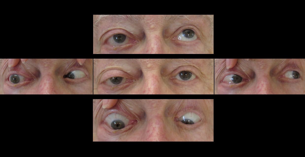
Figure 1. Extraocular motility at presentation.
Neuroimaging was obtained. Brain and orbits magnetic resonance imaging (MRI) with and without contrast (Figure 2) showed abnormal thickening and enlargement of multiple EOMs including the right medial rectus, right lateral rectus, right superior oblique, and right superior rectus. In the setting of the patient’s known lymphoma, this clinical picture was concerning for lymphomatous involvement of the EOMs and orbit. A positron emission tomography (PET) / computed tomography (CT) study (Figure 3) was then performed, which showed new abnormal fluorodeoxyglucose (FDG) uptake in the right optic nerve and right extraocular muscles as well as in several other areas including the right pre-maxillary tissue. The right eye proptosis was also apparent on axial cuts. The right pre-maxillary tissue was determined to be most accessible for biopsy and to carry the least morbidity. The biopsy indeed showed B-lymphoproliferative disease that was consistent with the patient’s known diffuse large B-cell lymphoma.
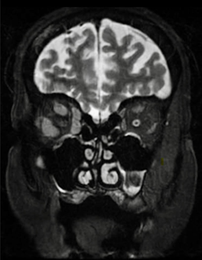
Figure 2. Orbital T2 MRI with fat suppression demonstrating enlarged EOMs in the right orbit.
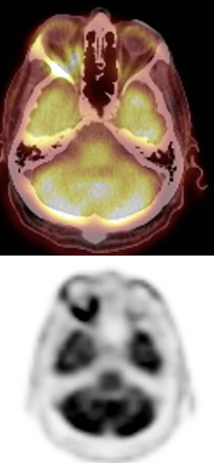
Figure 3. PET CT demonstrating abnormal FDG uptake within the right optic nerve and the right EOMs. Also note right eye proptosis.
Given the guarded systemic prognosis and after extensive discussion with the patient, the medical team, and the patient’s family, the goals of care ultimately became comfort. He was started on systemic corticosteroids, which improved his ocular motility (Figure 4). Moisture chamber goggles were initiated to protect the right eye given the extent of his proptosis and risk of cornea and conjunctival exposure. Regular ophthalmic lubrication was also recommended. The patient opted for a black occluding patch to eliminate his binocular diplopia. The patient passed away about one month after his initial presentation.
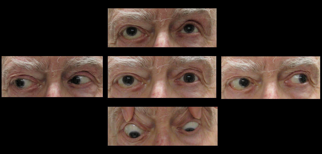
Figure 4. Extraocular motility following systemic steroids.
Discussion
Orbital disease may be a kind of “blind spot” for eye care providers. We are used to being able to directly look at a structure and diagnose a condition (e.g. examine the crystalline lens, diagnose a cataract). However, the orbit is not readily visualized during the clinical exam, and ancillary clues are necessary in order to arouse suspicion of an orbital process. Orbital disease presents in many ways. In this patient, proptosis, restricted eye movements, headache/pressure sensation, and chemosis were helpful clinical signs suggesting an orbital process. Notably, afferent dysfunction (i.e., reduced visual acuity, abnormal pupil reactivity, dyschromatopsia, and reduced brightness sense) also suggested optic nerve dysfunction, which was later confirmed on PET/CT imaging, which showed lymphomatous optic nerve involvement.
Orbital lymphoid malignancies are increasingly recognized as a significant cause of disability in the realm of orbital disease. Lymphoma has the propensity to mold to surrounding structures, erode bone, and infiltrate tissue. These cancers and other benign lymphoproliferative disorders are notoriously heterogeneous, with many possible clinical presentations.1,2,3 Importantly, benign and malignant forms of lymphoproliferative disease cannot be reliably differentiated by radiology (e.g., CT or MRI) alone, and biopsy of affected tissue is usually required.3,4 Among lymphoid malignancies, there are several subtypes including mucosa-associated lymphoid tissue (MALT) lymphomas (extra-nodal marginal zone lymphoma) vs non-MALT lymphomas.2 The patient in our case report had diffuse large B-cell lymphoma (DLBCL), a form of non-Hodgkin lymphoma that can be aggressive. Treatment may involve radiation, chemoimmunotherapy, and/or surgery. Observation may be considered. Systemic corticosteroids can shrink or lyse lymphomatous lesions, which resulted in improved extraocular motility in this patient’s case. However, biopsy is preferred prior to steroid initiation in order to maximize diagnostic yield of the tissue sampling, as lymphomas can sometimes undergo a transformation from an indolent to aggressive subtype.4,5
Management of orbital lymphoma mandates an interdisciplinary approach. Eye care, hematology/oncology, endocrinology, social work, palliative care, nutrition specialists, and other medical specialists all worked together to help manage this patient. Care, attention, and emotional support were also provided to the patient’s family throughout his care.
Clinical Pearls
- Eye care providers must recognize proptosis, chemosis, and ocular motility restriction as possible indicators of orbital disease
- Orbital lymphoma should be considered in the differential diagnosis for new proptosis in susceptible individuals
- As with most forms of cancer, orbital lymphoma requires an inter-disciplinary approach, and eye care providers must be aware of the potential for ophthalmic complications
References
- Shinder R, Al-Zubidi N, Esmaeli B. Survey of orbital tumors at a comprehensive cancer center in the United States. Head Neck. 2011;33:610-614. https://doi.org/10.1002/hed.21498
- Peres C, et al. Orbital cancer: Historical trends in histological composition and cause-specific mortality based on SEER database analysis. J Clin Oncol. 2026;44:e18138-e18138. doi:10.1200/JCO.2026.44.16_suppl.e18138
- Wang X, Zhang M, Chen Y, Wang X, Cui H. Characteristics of orbital lymphoproliferative disorders: a retrospective study of 84 cases. Eye (Lond). 2025;39(10):2096-2103. doi:10.1038/s41433-025-03816-z
- Mombaerts I, Rose GE, Garrity JA. Orbital inflammation: Biopsy first. Surv Ophthalmol. 2016;61(5):664-669. doi:10.1016/j.survophthal.2016.03.002
- Plotkin SR, Batchelor TT. Primary nervous-system lymphoma. Lancet Oncol. 2001;2(6):354-365. doi:10.1016/S1470-2045(00)00390-9

Dr. Kane graduated from New England College of Optometry in 2015 and went on to complete an ocular disease/primary care residency at VA Boston Jamaica Plain from 2015-2016. He is currently an attending optometrist at VA Boston. His interests include clinical teaching, neuro-ophthalmic disease, retinal vascular disease, glaucoma, and ocular manifestations of systemic disease.

Dr. Michelle Curtis earned her Doctor of Optometry degree from the New England College of Optometry in 2025. She subsequently completed an ocular disease residency at the VA Boston Healthcare System in West Roxbury, gaining advanced training in the diagnosis and management of complex ocular disease and systemic conditions with ocular manifestations. Dr. Curtis currently practices in Somerville, Massachusetts, with a clinical focus on comprehensive eye care and ocular disease management.

Daniel R. Lefebvre, MD, FACS, is an oculoplastic and orbital surgeon, Assistant Professor of Ophthalmology at Harvard Medical School, Mass Eye and Ear, member of the American Society of Ophthalmic Plastic and Reconstructive Surgeon (ASOPRS), and a staff surgeon at the Veterans Administration Boston HealthCare System in Boston, MA.