


Central Corneal Pseudomicrocysts Resultant from Belantamab Mafodotin Therapy: A Case Report
 Save as PDF
Conflict of interest: The author(s) declared that this work was conducted in the absence of any commercial or financial relationships that could be construed as a potential conflict of interest.
Abstract
Background: Multiple myeloma is characterized by uncontrolled plasma cell growth in bone marrow, ultimately leading to significant bone destruction. Belantamab mafodotin is an antibody drug conjugate approved for the treatment of patients with heavily pretreated relapsed or refractory multiple myeloma (RRMM). Despite its therapeutic benefits, belantamab mafodotin has been associated with significant ocular adverse effects, most notably microcystic epithelial changes, or corneal pseudomicrocysts.
Case Report: A 65-year-old White male presented to the clinic with blurry vision, right eye worse than left eye, that started one month prior to presentation. On ocular examination, the patient’s best corrected visual acuity was 20/30 in the right eye and 20/20 in the left eye; previously, it had been 20/20 in each eye. Entrance testing, including confrontation visual fields, extraocular muscle movement, and pupils, was all within normal limits. Slit lamp microscopy examination was most notable for an infero-central sub-epithelial haze in both eyes with an isolated 1.5 mm epithelial cyst in the right eye. Anterior segment optical coherence tomography showed a raised, superficial hyper-reflective epithelial pseudomicrocyst.
Conclusion: Belantamab mafodotin remains an important therapeutic option for patients with RRMM. However, corneal pseudomicrocysts can develop and represent a common form of ocular toxicity that may require dose modification or cessation. This case demonstrates the importance of routine ocular monitoring and emphasizes the need for interdisciplinary collaboration and communication among all health care providers to optimize patient care and treatment outcomes for patients with RRMM treated with belantamab mafodotin.
Keywords: belantamab mafodotin, cornea, microcystic epithelial changes, pseudomicrocysts
Introduction
Multiple myeloma is the second most prevalent hematological malignancy that accounts for about 1% of all cancers.1 It is characterized by uncontrolled plasma cell growth in bone marrow, leading to significant bone destruction. Belantamab mafodotin is a new antibody drug conjugate (ADC) approved for the treatment for patients with heavily pretreated relapsed or refractory multiple myeloma (RRMM). Despite its therapeutic efficacy, this treatment is often associated with ocular toxicity, most notably corneal pseudomicrocysts.
Belantamab mafodotin is composed of anti-B cell maturation antigens (BCMA), a tumor necrosis factor surface receptor, that is linked to microtubule inhibitor monometyl auristatin F (MMAF), a toxic drug molecule found in ADCs.1 Once belantamab mafodotin is internalized, MMAF is released intracellularly, resulting in apoptosis of targeted malignant plasma cells.1 However, off-target uptake of MMAF by corneal epithelial cells can occur, leading to the development of pseudomicrocysts. These often present as bilateral, superficial cyst-like lesions and often require dose modifications based on severity. Given the high incidence of ocular toxicity, close communication and collaboration among eye care providers, oncologists, and other members of the patient’s healthcare team is essential to ensure appropriate management. This article will discuss a patient with corneal changes secondary to belantamab mafodotin.
Case Report
A 65-year-old White male presented to the clinic with blurry vision, right eye worse than left eye, that started one month prior to presentation. The patient’s ocular history was notable for age-related cataracts, dry eye, and history of diplopia secondary to a cranial nerve six palsy. The patient’s medical history was significant for post-traumatic stress disorder (PTSD), anxiety, depression, sleep apnea, hyperlipidemia, vitamin D deficiency, vitamin B12 deficiency, osteoarthritis, and multiple myeloma. Current medications included baby aspirin, cyanocobalamin, belantamab mafodotin, preservative-free carboxymethylcellulose solution, and cyclosporine 0.05% ophthalmic solution.
Upon further questioning, the patient acknowledged that the blurred vision was likely an adverse effect of his current multiple myeloma treatment, as he had been previously informed that this commonly occurs following each administration. The patient reported a history of fluctuating visual symptoms following treatment but indicated that the current presentation was more severe than any prior episode. The patient was routinely followed by a corneal specialist who works closely with his oncologist; however, he was unable to secure an appointment in a timely manner.
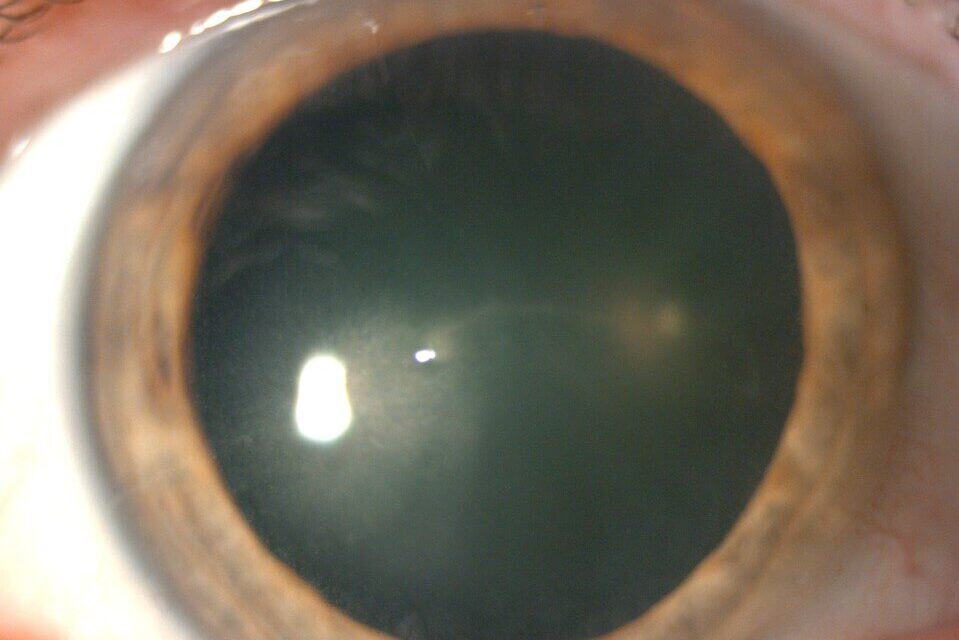
Upon ocular examination, the patient’s best corrected visual acuity (BCVA) was 20/30 in the right eye and 20/20 in the left eye, previously 20/20 in the right and left eye. All entrance testing, including confrontation visual fields, extraocular muscle movement, and pupils were all within normal limits. Slit lamp microscopy examination revealed 1+ meibomian gland dysfunction in both eyes, 2+ diffuse superior punctate epithelial erosions (SPEE) in both eyes, and an infero-central sub-epithelial haze in both eyes with an isolated 1.5 mm epithelial cyst in the right eye. Anterior segment optical coherence tomography (AS-OCT) was performed, which showed a raised, superficial, hyperreflective epithelial pseudocyst.
Six years prior to his presentation, the patient had been symptomatic for right-side rib pain for several weeks and hip pain for the prior one or two years. Chest computed tomography (CT) revealed a 10.4 cm dominant soft tissue mass with complete destruction of the left posterior seventh rib. X-rays of the hip showed calcifications on the right trochanter suggestive of calcific tendinitis and degenerative narrowing of the right hip. He was later officially diagnosed with Immunoglobulin G (IgG) kappa multiple myeloma and started on Bortezomib, lenalidomide, and dexamethasone combination therapy. Over the years, the patient had tried a variety of therapies, such as Cytoxan and Selinexor. Nine months prior to the patient’s presentation to the clinic, Blenrep (belantamab mafodotin) treatment was initiated for the patient.
The patient was evaluated by his corneal specialist three weeks after the onset of corneal symptoms, where he was instructed to begin artificial tears every two to four hours, gel artificial tears every night, and Muro 128 every night. The corneal specialist communicated with the patient’s oncologist and recommended withholding the next dose of therapy pending further ocular evaluation. The patient was further advised to continue close follow-up with his oncology team.
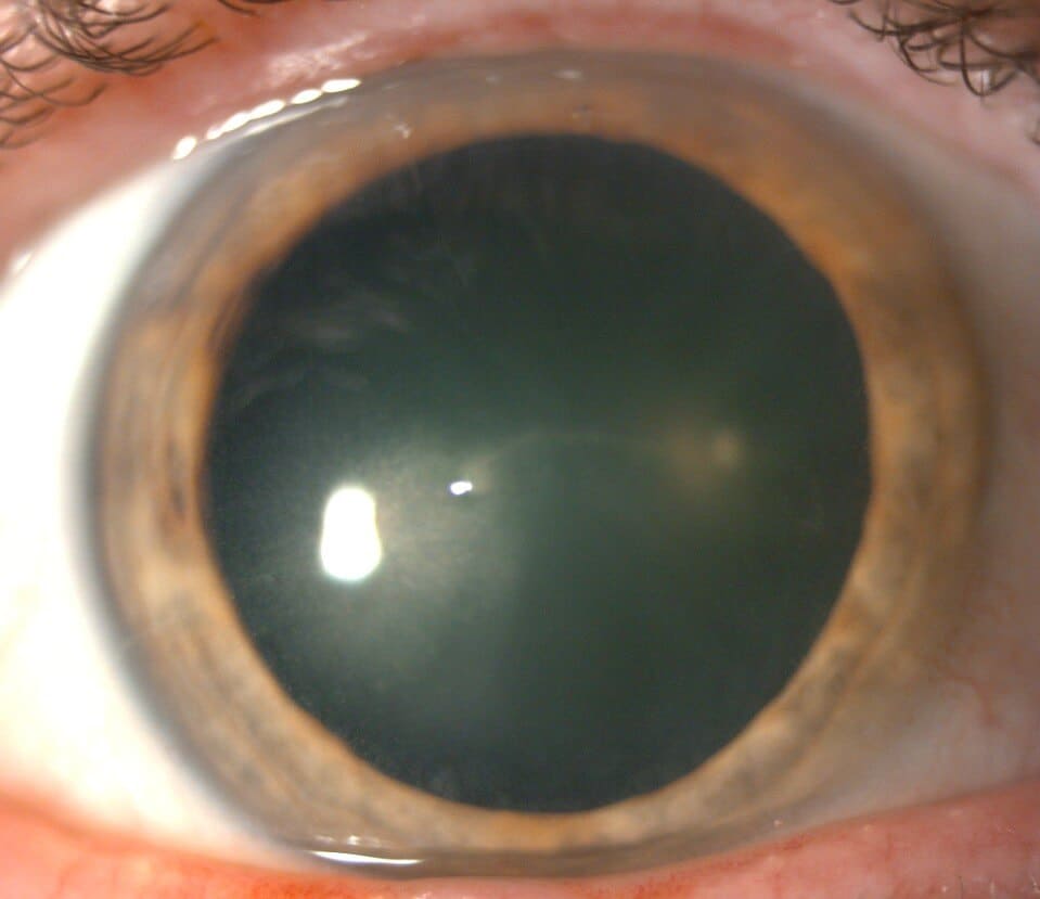
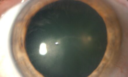
Figure 1: This slit lamp photo of the right eye shows mild epithelial haze just inferior nasal to the visual axis.

Figure 2: This anterior segment OCT cross section highlights the hyper-reflective and raised pseudocyst.
Discussion
Multiple myeloma is a neoplastic proliferation of plasma cells that produce monoclonal immunoglobulin. These plasma cells proliferate within the bone and cause skeletal destruction, such as osteolytic lesions, osteopenia, and pathologic fractures.2 Primary refractory multiple myeloma occurs when the patient fails to obtain a minimal response on initial therapy and progresses on current therapy. Relapsed or refractory multiple myeloma occurs when there is progressive disease, poor response despite treatment, progression within 60 days of the most recent treatment in a patient who had achieved remission, the absence of minimal response or primary refractory multiple myeloma.3
Belantamab mafodotin was initially approved by the United States Food and Drug Administration (FDA) and the European Medicines Agency (EMA) in August 2020, for the treatment of adults with RRMM who have received at least four prior therapies, including an anti-CD38 monoclonal antibody, a proteasome inhibitor, and an immunomodulatory agent.4 However, in the phase 3 DREAMM 3 trial, belantamab mafodotin monotherapy did not meet its primary endpoint of progression-free survival (PFS), defined as the duration during and after receiving treatment that the patient lives without disease progression, compared to pomalidomide with dexamethasone in patients with RRMM.5 As a result, belantamab mafodotin was withdrawn from the US, the European Union, and the United Kingdom in 2024.5
The DREAMM clinical trial program evaluates the efficacy and safety of belantamab mafodotin monotherapy in comparison with current treatment regiments. The DREAMM-7 trial in phase 3 of the study further evaluated the combination of belantamab mafodotin, bortezomib, and dexamethasone (BVd) with the combination of daratumumab, bortezomib, and dexamethasone (DVd) in patients with RRMM with more than one prior line of therapy.6 It was concluded that there was benefit in the PFS and overall survival in BVd group. After the completion of the DREAMM-7 trial, belantamab mafodotin was officially FDA-approved on October 23, 2025, for the treatment of adults with RRMM who have received at least two prior lines of therapy.6
Belantamab mafodotin is an antibody drug conjugate (ADC), consisting of monoclonal antibodies linked to a cytotoxic cell surface receptor by a chemical linker. The cell surface receptor is a highly potent cytotoxic payload.7 The relationship between the cytotoxic payload and the antibody contributes to the effectiveness of the ADC. This three-component combination delivers cytotoxic agents to tumor sites by antigen recognition, while sparing healthy tissues by off-target effects.7 Belantamab mafodotin comprises of an IgG1 monoclonal antibody that targets Anti-B-Cell Maturation Antigen (BCMA).1 BCMA is a tumor necrosis factor surface receptor found on malignant plasma cells but absent on normal cells.8 BCMA is linked to microtubule inhibitor monomethyl auristatin F (MMAF), the cytotoxic payload found in all ADCs.1 Upon binding to BCMA, belantamab mafodotin is internalized by endocytosis and transported to lysosomes, where MMAF is released in the acidotic intracellular environment.1 This results in a target therapy that induces apoptosis and triggers immunogenic cell death, and immunotherapy that enhances antibody-dependent cellular toxicity and phagocytosis.
Belantamab mafodotin, along with other ADCs, has been associated with significant ocular adverse reactions. In the phase 2 DREAMM-2 trial, patients underwent corneal examinations every three weeks by an optometrist or ophthalmologist. Ocular examinations included BCVA and a thorough slit lamp examination.9 The most common adverse events with belantamab mafodotin, as outlined in the DREAMM-2 study, included keratopathy (72%), thrombocytopenia (38%), and infusion-related reactions (21%).8 The most common keratopathy noted were microcystic epithelial changes (MECs), also known as corneal pseudomicrocysts. These lesions are typically bilateral, superficial, micro-cystic like lesions that can present with sub-epithelial haze in the moderate to severe categories.8 These MECs begin centrally and migrate peripherally. Correspondingly, blurred vision and dry eye were the most common symptoms associated with belantamab mafodotin use.
The ocular adverse events observed with belantamab mafodotin are attributed to off-target toxicity. This is primarily due to microtubule-disrupting monomethyl auristatin F (MMAF), the cytotoxic component linked to the monoclonal antibody via a protease-resistant maleimidocaproyl (mc) linker. Off-target damage may result from linker instability, premature extracellular cleavage, intracellular metabolism of the linker-cytotoxin complex, or Fc-receptor mediated cellular uptakes.10 In corneal cells, MMAF inhibits basal epithelial cell division. Once internalized into the basal epithelial layer, it disrupts cell proliferation and induces apoptosis. These apoptotic cells are displaced anteriorly, forming the pseudomicrocysts.10
On average, MECs were observed at a median time of 37 days.8 Approximately 25% of patients developed MECs after the first dose, increasing to 69% by the fourth dose.9 Based on the severity of the corneal findings, modifications are often necessary. The Keratopathy and Visual Acuity (KVA) scale was developed during the DREAMM 2 trial to guide dosing modifications based on severity of MEC’s.8
- Grade 1 (mild): majority in periphery (>80%), BCVA one line worse than baseline
- Grade 2 (moderate): predominantly paracentral, BCVA 2-3 lines worse than baseline
- Grade 3 (severe): central, 3 or more lines worse than baseline (<20/200)
- Grade 4 (very severe): central, BCVA worse than 20/200
Grading is based on the worst finding, either BCVA or lesion location. Grade 3 and 4 MECs were most commonly observed, accounting for 45% of cases.9 Given this patient’s decreased BCVA (20/30) and predominantly paracentral location of the corneal pseudo microcyst, these findings were consistent with a grade 2 toxicity. The severity of MECs frequently leads to dose modification, accounting for 47% of dose delays and 25% of dose reductions.9 For grade 1 findings, continuation of the current belantamab mafodotin dose is appropriate.11 In grade 2 and 3 toxicity, treatment should be delayed until the pseudomicrocysts improve to a grade 1 or fully resolve.11 For grade 4 toxicity, a thorough benefit-risk assessment is required to determine whether treatment discontinuation is appropriate.11 Given the patient’s grade 2 pseudomicrocysts, treatment was held until the ocular toxicity was resolved. The most common symptoms associated with corneal pseudomicrocysts include blurred vision and dry eye.9 Because these adverse effects are dose-dependent, management primarily involves supportive therapy with artificial tears for lubrication.11
It is important to note that clinical findings do not always correlate with patient-reported symptoms or changes in BCVA. In the DREAMM-2 trial, only 56% of patients with MECs reported symptoms such as dry eye and blurred vision, and/or had a decrease in BCVA (≥ 2 lines decline in the better-seeing eye).8
Given the severity and frequency of the corneal pseudomicrocysts, coordinated care among all healthcare providers is essential. The current recommended dosage of belantamab mafodotin is 2.5 mg/kg. It is administered via infusions once every three weeks in conjunction with bortezomib and dexamethasone for the first eight cycles, followed by monotherapy until disease progression or unacceptable toxicity occurs.6 Eye exams are required at baseline before the initial administration of belantamab mafodotin, and before cycles 2, 3, and 4.12 Per protocol, all MECs must be followed by the eye care provider until full resolution or recovery to baseline.9 After the fourth cycle of administration, the physician may use the Vision-Related Anamnestic (VRA) tool, a questionnaire assessing patient-reported ocular symptoms and its impacts on daily activities.12 This questionnaire, outlined below, is used to determine if the patient qualifies to receive the next dose of belantamab mafodotin or to refer to an eye care provider for an eye examination.12 If the patient answers “Yes” to any question, and the ocular event or its impact on activities of daily living are worsening and the symptoms are persisting for more than eight hours, then belantamab administration should be delayed until the adverse ocular event has resolved.12
- Question 1: During the last 24h, did you ever feel that your eyes were sensitive to light?
- Question 2: During the last 24h, did your eyes ever feel gritty?
- Question 3: During the last 24h, did your eyes feel painful or sore?
- Question 4: During the last 24h, did you ever experience blurred vision?
- Question 5: During the last 24h, did you ever experience poor vision?
- Question 6: During the last 24h, did you ever experience problems in reading due to problems with your eyes?
- Question 7: During the last 24h, did you ever experience problems in driving due to problems with your eyes?
- Question 8: During the last 24h, did you ever find it difficult to work with a computer or a smartphone due to problems with your eyes?
- Question 9: During the last 24h, did you ever find it difficult to watch TV due to problems with your eyes?
Physicians should work and communicate closely with eye care providers throughout the entire administration of belantamab mafodotin, especially to assess Grade 4 ocular events. Though these pseudomicrocysts eventually resolve with dose cessation or modifications, risk-benefit assessment should be conducted to determine whether to hold off on treatment or continue treatment despite risks.12 It is evident that communication between eye care providers, hematologists, and oncologists is imperative throughout the treatment course for these patients. Eye care providers must perform thorough ocular examinations and report back to patients’ hematologists or oncologists, who can ultimately determine dosing modifications.
Conclusion
Belantamab mafodotin represents an important advancement in the treatment of relapsed or refractory multiple myeloma. Its mechanism as an ADC targeted to tumor cells allows for effective antitumor therapy. However, it is accompanied by significant risk of ocular toxicity. Corneal pseudomicrocysts are dose-dependent and can significantly impact visual outcomes. The implementation of the KVA scale helped establish guidelines for monitoring and managing these ocular findings based on BCVA and the location of the pseudomicrocysts.
Ultimately, the successful use of belantamab mafodotin relies on a multidisciplinary approach. This case highlights the importance of interdisciplinary care and effective communication among health care providers. Close collaboration between eye care providers, hematologists, and oncologists is essential to ensure proper patient-centered decision making. Through coordinated multidisciplinary management, both visual and overall systemic health outcomes may be optimized in this patient population.
References
- Almodovar Diaz AA, Alouch SS, Chawla Y, Gonsalves WI. The Antibody Drug Conjugate, Belantamab-Mafodotin, in the Treatment of Multiple Myeloma: A Comprehensive Review. Blood Lymphat Cancer. 2024;14:71-87. Published 2024 Dec 6. doi:10.2147/BLCTT.S490021
- Laubach, Jacob. Multiple myeloma: Clinical features, laboratory manifestations, and diagnosis. In: UpToDate, Connor RF (Ed), Wolters Kluwer. (Accessed on April 18, 2026)
- Ahmed A, Killeen RB. Relapsed and Refractory Multiple Myeloma. [Updated 2023 Jun 8]. In: StatPearls [Internet]. Treasure Island (FL): StatPearls Publishing; 2026 Jan. https://www.ncbi.nlm.nih.gov/books/NBK592405/
- Baines AC, Ershler R, Kanapuru B, et al. FDA Approval Summary: Belantamab Mafodotin for Patients with Relapsed or Refractory Multiple Myeloma. Clin Cancer Res. 2022;28(21):4629-4633. doi:10.1158/1078-0432.CCR-22-0618
- Mukhopadhyay P, Abdullah HA, Opalinska JB, et al. The clinical journey of belantamab mafodotin in relapsed or refractory multiple myeloma: lessons in drug development. Blood Cancer J. 15, 15 (2025). https://doi.org/10.1038/s41408-025-01212-0
- Center for Drug Evaluation and Research. FDA approves belantamab Mafodotin-blmf for relapsed or refractory multiple myeloma. U.S. Food and Drug Administration. October 23, 2025. Accessed April 18, 2026. https://www.fda.gov/drugs/resources-information-approved-drugs/fda-approves-belantamab-mafodotin-blmf-relapsed-or-refractory-multiple-myeloma
- Wang R, Hu B, Pan Z, et al. Antibody-Drug Conjugates (ADCs): current and future biopharmaceuticals. J Hematol Oncol. 2025;18(1):51. Published 2025 Apr 30. doi:10.1186/s13045-025-01704-3
- Lonial S, Nooka AK, Thulasi P, et al. Management of belantamab mafodotin-associated corneal events in patients with relapsed or refractory multiple myeloma (RRMM). Blood Cancer J. 2021;11:103. doi:10.1038/s41408-021-00494-4
- Farooq AV, Degli Esposti S, Popat R, et al. Corneal Epithelial Findings in Patients with Multiple Myeloma Treated with Antibody-Drug Conjugate Belantamab Mafodotin in the Pivotal, Randomized, DREAMM-2 Study. Ophthalmol Ther. 2020;9(4):889-911. doi:10.1007/s40123-020-00280-8
- Wahab A, Rafae A, Mushtaq K, et al. Ocular Toxicity of Belantamab Mafodotin, an Oncological Perspective of Management in Relapsed and Refractory Multiple Myeloma. Front Oncol. 2021;11:678634. Published 2021 May 11. doi:10.3389/fonc.2021.678634
- Arazi M, Wattad A, Magen H, et al. Interdisciplinary management of belantamab mafodotin-associated ocular toxicity in clinical practice. Clinicalkey. Published October 1, 2025. Accessed May 1, 2026. https://doi.org/10.1016/j.jtos.2025.09.003
- Terpos E, Trudel S, Mateos MV, et al. Practical Guidance on Clinical Management of Belantamab Mafodotin-Associated Ocular Events. Am J Hematol. 2025;100(10):1839-1850. doi:10.1002/ajh.70015

Dr. Wang graduated from the New England College of Optometry in 2025. She completed her ocular disease residency at the VA Boston Healthcare System.