


Post-operative Hypotony And Choroidal Effusion Following Cataract Surgery Suture Removal

 Save as PDF
ABSTRACT
BACKGROUND
Ocular hypotony can occur for a variety of reasons, including but not limited to ocular inflammation, glaucoma filtration surgery, or trauma. This case report describes ocular hypotony following premature corneal suture removal after cataract surgery. This case also provides a detailed account of the ocular complications associated with ocular hypotony – including choroidal effusion and choroidal folds – and the risk factors and appropriate treatment options for these complications.
CASE REPORT
An 80-year-old Caucasian male presented as an urgent visit due to new onset blurry vision and dull eye pain several days after corneal suture removal after cataract surgery. Initial testing revealed a low intraocular pressure and positive Seidel sign at the wound site. Furthermore, dilated fundus exam showed areas of choroidal effusion and choroidal folds, both complications of ocular hypotony. The patient was immediately sent to his surgeon to close the wound and reform the anterior chamber.
CONCLUSION
Due to the various causes of ocular hypotony, a thorough case history and careful ocular evaluation are imperative to determine the underlying cause and potential vision-threatening complications. Understanding the cause and complications allows for appropriate treatment and referral, if necessary, potentially minimizing the risks of long-term negative impacts of ocular hypotony.
Keywords: hypotony, choroidal effusion, choroidal folds, wound leak, suture removal, post-operative cataract surgery
INTRODUCTION
Ocular hypotony is most commonly described as intraocular pressure (IOP) lower than 5mmHg,1 but has also been reported as any IOP lower than 10mmHg.2 While ocular hypotony most often occurs after glaucoma filtration surgery, other potential causes include trauma, inflammation, wound leaks, medications, or systemic conditions.1 Complications of ocular hypotony include choroidal effusion or detachment, choroidal folds, hypotony maculopathy, and refractive error changes.1 Urgent treatment is imperative for best visual recovery and is dependent on the causative agent. This case report highlights ocular hypotony and the associated complications with a unique cause of post-operative (post-op) cataract surgery corneal suture removal.
CASE REPORT
An 80-year-old Caucasian male presented with a complaint of blurry, foggy vision OD with dull, intermittent pain and soreness in the lower right corner and upper cheek that radiated behind the eye. The patient denied trauma or aggressive eye rubbing. Ten days prior to appointment, the patient had cataract surgery OD which required a Malyugin ring and three nylon sutures at the incision site. The site was found to be watertight both immediately after the procedure, and immediately after the suture removal at the recent one-week post-op visit.
The patient’s medical history was positive for a history of stroke, coronary artery disease, and lower extremity deep venous thrombosis. The patient’s systemic medications were ascorbic acid, coenzyme Q10, pyridoxine hydrochloride, turmeric, and cyanocobalamin. He was using post-cataract surgery topical medications: prednisolone acetate 1% QID OD, moxifloxacin 0.5% QID OD, and ketorolac 0.5% QID OD.
The patient presented with uncorrected distance vision OD 20/150. Pertinent corneal slit lamp findings OD revealed 3+ endothelial folds, 2+ temporal edema, iridocorneal touch inferior temporal with overlying epithelial heaping, the iris extending anteriorly to the temporal corneal incision site, and a positive Seidel sign at the incision site. The anterior chamber OD was shallow with trace cells and free-floating iris inferior temporal into the anterior chamber with an iris blood vessel extending from the inferior temporal incision site of cornea. The iris OD showed no evidence of iris prolapse.
IOP was measured with multiple instruments. Ocular response analyzer (ORA) revealed IOP of 1.0mmHg OD and 10mmHg OS. iCare tonometry was attempted OD without success (the instrument could not obtain a reading) and measured 10mmHg OS. Goldman applanation tonometry was performed with results of 0mmHg OD and 10mmHg OS.
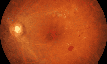
With verbal consent, the patient was dilated OU with one drop 1% tropicamide and one drop 2.5% phenylephrine. Posterior segment findings OD revealed a centered posterior chamber intraocular lens (PCIOL) with 2-3+ diffuse posterior capsular opacification (PCO) and capsular fibrosis. The vitreous revealed syneresis but was clear of inflammation. The optic nerve, vessels, and macula were unremarkable, with no apparent macular edema. The mid-peripheral and peripheral retina revealed inferior, inferior temporal, and inferior nasal choroidal effusion (Figure 1 A-E). No holes, tears, or retinal detachment were present. Choroidal folds were present throughout the posterior pole OD.

Figure 1A: Photo montage of OD fundus on day 1

Figure 1B: OD nasal fundus showing choroidal effusion

Figure 1C: OD inferior fundus showing choroidal effusion

Figure 1D: OD temporal fundus showing choroidal effusion

Figure 1E: OD posterior pole showing choroidal effusion
A macula optical coherence tomography (OCT) was performed to confirm and further assess the subtle choroidal folds OD (Figure 2) with no other significant findings noted.

Figure 2. Right eye macula OCT B-scans on initial presentation through various cross sections demonstrating choroidal folds.
The patient was urgently referred to his surgeon for corneal wound leak repair due to ocular hypotony with positive Seidel after suture removal. The surgeon recommended immediate anterior chamber washout with iris repositioning and sutured wound revision OD. A bandage contact lens with protective eye shield was placed on OD until the surgeon arrived in clinic to perform the repair.
Post-Op Wound Revision Day 1
The patient reported with uncorrected distance vision OD 20/200. IOP with Goldman applanation tonometry was 6 mmHg OD. Pertinent slit lamp findings OD revealed 3-4+ temporal corneal edema at the keratome incision site with 3 interrupted sutures, Seidel sign negative at the wound, and 1-2+ central corneal edema. The anterior chamber was deep with 1-2 rare cells and trace flare. The iris had mild temporal atrophy near the wound repair with no neovascularization. Posterior exam findings were similar to initial presentation with slight improvement in choroidal effusion (Figure 3) and choroidal folds (Figure 4).

Figure 3A: Photo montage of OD fundus on post-op day 1

Figure 3B: OD nasal fundus showing improvement in choroidal effusion

Figure 3C: OD inferior fundus showing improvement in choroidal effusion

Figure 3D: OD temporal fundus showing improvement in choroidal effusion

Figure 3E: OD posterior pole showing improvement in choroidal effusion

Figure 4. Right eye macula OCT on post-op revision day 1 through various cross sections showing improvement of choroidal folds.
The plan was to continue prednisolone acetate 1% QID OD, moxifloxacin 0.5% QID OD, and wear an eye shield every night at bedtime OD. A follow-up was scheduled for three days.
Post-Op Wound Revision Day 45
The patient was seen at two-week intervals before proceeding with suture removal. During these visits, visual acuity, IOP, clinical findings, and patient symptoms were similar to post-op revision day 1.
On day 45 follow-up, corneal sutures were removed, and the patient stayed in the clinic for one hour to ensure the wound did not start re-leaking. Pertinent slit lamp findings OD revealed 1+ corneal edema and negative Seidel sign at the temporal keratome incision site, indicating that the wound was well sealed. The anterior chamber OD was deep and quiet, and there was temporal iris atrophy. Due to a well-sealed wound and negative Seidel sign, the patient was released and planned to follow up for a final visit in one week to ensure appropriate healing.
Final Post-op Wound Revision Day 52
Pertinent slit lamp findings OD revealed corneal stromal scarring at the temporal keratome incision, determined to be sealed with negative Seidel sign. Posterior segment findings OD were unremarkable with complete resolution of choroidal effusion and choroidal folds.
Due to complete resolution of ocular hypotony and retinal findings, the patient was released from care and sent for cataract surgery on the fellow eye.
DISCUSSION
Hypotony is defined as low IOP with numerical values that vary greatly from study to study. According to Wang et. al in Ocular Hypotony: A Comprehensive Review, ocular hypotony is defined as “…a numerical cutoff of ≤ 5 mmHg.”9 The American Academy of Ophthalmology defines hypotony as IOP less than 6.5 mmHg.10 The most common definition is an IOP lower than 5 mmHg. Ocular hypotony occurs due to an abundance of outflow of aqueous humor or insufficient aqueous humor production,9 both of which result in a drastic decrease in IOP.
Risk factors of ocular hypotony include surgery, trauma, or intraocular inflammation, and can be caused by a variety of primary or secondary conditions, the most common being leakage through ocular surgical wounds (as in this case report), reduction in aqueous humor production, topical or systemic medications, and proliferative vitreoretinopathy.1
Ocular hypotony following cataract surgery is due to excessive outflow of aqueous humor through the surgical incision site and can occur in patients with or without sutures.9 In this case report, the nylon sutures were removed from the incision site prematurely without proper corneal healing time, causing leakage of aqueous humor out of the incision site and resultant ocular hypotony.
Patients with ocular hypotony may be asymptomatic; therefore, diagnosis requires a thorough case history to determine recent or past ocular surgeries, trauma, inflammation, medications, or systemic conditions. Symptoms include vision loss, visual distortions, mild pain, photophobia, or general discomfort.9 If longstanding and chronic, patients may have complete vision loss due to complications.
Clinical signs of ocular hypotony include IOP lower than 5 mmHg, a perforated globe, or bleb or wound leak if post-surgical.1 Seidel testing with sodium fluorescein is imperative to confirm or refute a wound leak. Anterior segment findings may include corneal edema, shallow anterior chamber, or phacodonesis.1 Inflammation may or may not be present and could include corneal endothelium keratic precipitates or synechiae.1 Posterior segment findings include choroidal detachment, retinal hemorrhages, tortuous blood vessels, optic nerve edema, choroidal folds, or retinal pigment epithelial changes in the macula.1 In advanced cases, rhegmatogenous retinal detachment, proliferative vitreoretinopathy, or ocular ischemia may be seen.1
Differential diagnosis of ocular hypotony include ruptured globe, phthisis bulbi, retinal detachment, iridocyclitis, severe dehydration, cyclodialysis cleft, ocular ischemia, medications (i.e., ocular hypotensives), postoperative, or traumatic ciliary body shutdown.1,9
The complications associated with ocular hypotony contribute to symptoms which ultimately prompt patients to seek care, as seen in the patient in this case report. Complications can include refractive error shifts, corneal edema, maculopathy, choroidal effusion, or choroidal folds.1
Both hyperopic or myopic refractive error changes can occur from ocular hypotony.1 Hyperopia occurs due to reduced axial diameter and myopia occurs due to forward displacement of the lens.1 Resolution of both occur spontaneously without permanent significance with treatment and resolution of the initial cause of hypotony.1 Corneal edema due to hypotony is thought to occur from reduced function of the endothelial pumps that normally function to pull fluid into the aqueous.10
Hypotony maculopathy occurs in up to 20% of cases, and most commonly occurs after glaucoma filtration surgery.1 Maculopathy occurs when the axial length decreases from surgery, creating retinal-choroidal folds, which then decreases the diameter of the vitreous cavity, causing the retina to fold around the fovea.13 Patients are typically asymptomatic but can report distortions in central vision.13 Treatment is guided by first determining the causative agent.
Choroidal effusion occurs due to abnormal collection of fluid in the suprachoroidal space which thickens the choroid and can cause a choroidal detachment.11 The two types of choroidal effusion are serous and hemorrhagic.12 Serous effusion is due to serum leaking from choroidal blood vessels to the suprachoroidal space.12 Hemorrhagic effusion occurs after worsening serous effusion when blood leaks from a ruptured ciliary blood vessel into the suprachoroidal space.12
Risk factors for choroidal effusion include glaucoma filtration surgery (most common), hypotony, post-operative inflammation, reduced IOP before or after cataract surgery, antiplatelet or anticoagulant medications, pseudophakia, or post-operative wound leaks.12
Symptoms of choroidal effusion vary depending on location and cause, and often mirror angle closure due to anterior displacement of structures. If the effusion is peripheral, there may be little or no impact on vision.12 If the effusion extends into the posterior pole, patients may experience blur or distorted vision.12 For hemorrhagic choroidal effusion, symptoms are often more obvious and include decreased vision and pain, which can also cause headaches, nausea, or vomiting.12
Clinical signs of choroidal effusion include an elevated choroid with retinal pigmented epithelium changes that appear as “leopard spot fundus.”11 Choroidal elevation causes easier visualization of the ora serrata, often extending close to and into the posterior pole.11 The fundus appears brown due to choroidal thickening, with normal appearing overlying retinal vessels.11 If the effusion is serous, subretinal fluid will be present.12 If the effusion is hemorrhagic, blood will be present.12 The anterior chamber is typically shallow and can lead to angle closure.11 The vitreous is unremarkable unless caused by a retinal detachment, in which case brown pigmented cells will be present (Shafer’s sign).11
Differential diagnosis of serous effusion includes serous retinal detachment (RD), angle closure attack, or pupillary block.11 Serous RDs often appear “wrinkled” on fundoscopy, compared to a smooth appearance of choroidal effusion.11 Differential diagnosis of hemorrhagic effusion includes choroidal melanoma, choroiditis, posterior scleritis, or central serous retinopathy.1
Choroidal folds, subretinal undulations of dark and bright striae throughout the retina, are another complication of post-cataract surgery hypotony.14 The folds are usually horizontal but can be in any direction.14 Causes for folds include hypotony, trauma, globe compression, orbital mass, orbital inflammation, or idiopathic.14 Patients may be asymptomatic or have decreased vision or metamorphopsia.14 Signs include choroid undulations which can be enhanced on slit lamp with a red-free filter.14 Additional testing to confirm diagnosis and determine etiology includes fluorescein angiography, OCT, or ultrasound B scan.14
Treatment for ocular hypotony, choroidal effusion, and choroidal folds depends heavily on the cause, and the management will be to diagnose the inciting incident, repair damage to the eye, and the aim to restore vision.9 Patients are encouraged to start a topical cycloplegic, which deepens anterior chamber and corticosteroid, an anti-inflammatory agent and can increase IOP.1 Topical hypotensive agents should be avoided or discontinued to prevent further IOP decrease.9 In severe hypotony, viscoelastic material can be injected into the anterior chamber to prevent further collapse.9 The patient in this case had an extremely shallow anterior chamber with an obvious wound leak after surgical incision, requiring immediate anterior chamber reformation and closure of the wound site. In all cases, patients are encouraged to wear protective eye wear for trauma prevention until consultation with a surgical specialist.
The prognosis of ocular hypotony greatly depends on the cause of insult, severity of complications, and timeliness of treatment, which is why a thorough case history is imperative in the successful management of these patients. Once the cause has been successfully identified and treated, hypotony typically resolves without further permanent damage.1 If hypotony is chronic, however, a worst case scenario is the development of phthisis bulbi.13 Overall, patients have a positive prognosis with very little long-term impact on eyes and vision.
CONCLUSION
Ocular hypotony after suture removal from cataract surgery can lead to many complications that could ultimately result in loss of vision if not managed promptly and appropriately. Choroidal effusion and choroidal folds are two common findings in ocular hypotony, which typically occur after glaucoma surgery. But as seen in this case report, they can also occur from a wound leak after premature corneal suture removal. Timing is key to closing the wound leak, restoring the anterior chamber, and elevating the intraocular pressure to avoid permanent vision loss.
REFERENCES
- Okonkwo ON, Tripathy K. Ocular Hypotony. StatPearls [Internet]. 2022 Aug 23. Treasure Island (FL): StatPearls Publishing; 2022 Jan -. Available from: https://www.ncbi.nlm.nih.gov/books/NBK582144/
- Sabti K, Lindley SK, Mansour M, Discepola M. Uveal Effusion After Cataract Surgery: An Echographic Study. Ophthalmology [Internet]. 2001 Jan; 108(1):100-3. Available from: https://pubmed.ncbi.nlm.nih.gov/11150272/
- Rossi T, Romano MR, Iannetta D, Romano V, Gualdi L, D’Agostino I, Ripandelli G. Cataract Surgery Practice Patterns Worldwide: A Survey. BMJ Open Ophthalmology [Internet]. 2021 Jan 13;6(1):e000464. Available from: https://pubmed.ncbi.nlm.nih.gov/33501377/
- Balal S, Jbari AS, Nitiahpapand R, Cook E, Akhtar W, Din N, Sharma A. Management and Outcomes of the Small Pupil in Cataract Surgery: Iris Hooks, Malyugin Ring, or Phenylephrine?. Eye [Internet]. 2020 Nov 12. 35(10): 2714-2718. Available from: https://www.ncbi.nlm.nih.gov/pmc/articles/PMC8452752/
- Jhani V, Sharma N, Vajpayee RB. Management of Intraoperative Miosis During Pediatric Cataract Surgery Using Healon 5. Middle East African Journal of Ophthalmology [Internet]. 2011 Jan-Mar; 18(1): 55-57. Available from: https://www.ncbi.nlm.nih.gov/pmc/articles/PMC3085153/
- Malyugin B. Recent Advances in Small Pupil Cataract Surgery. Current Opinion in Ophthalmology [Internet]. 2018 Jan; 29(1):40-47. Available from: https://pubmed.ncbi.nlm.nih.gov/29059105/
- Matossian C, Makari S, Potvin R. Cataract Surgery and Methods of Wound Closure: A Review. Clinical Ophthalmology [Internet]. 2015 May 22; 9:921-928. Available from: https://www.ncbi.nlm.nih.gov/pmc/articles/PMC4447171/pdf/opth-9-921.pdf
- Jones L, Rostov AT. Postop Predicaments in Cataract Procedures. Review of Optometry [Internet]. 2014 Oct 15. Available from: https://www.reviewofoptometry.com/article/postop-predicaments-in-cataract-procedures
- Wang Q, Thau A, Levin AV, Lee D. Ocular Hypotony: A Comprehensive Review. Survey of Ophthalmology [Internet]. 2019 Sept-Oct;64(5):619-638. Available from: https://pubmed.ncbi.nlm.nih.gov/31029581/
- Sriram A, Tania Tai TY. Resolution of Chronic Corneal Edema After Surgical Treatment for Ocular Hypotony. Journal of Glaucoma [Internet]. 2017 Jun;26(6):e187-e189. Available from: https://pubmed.ncbi.nlm.nih.gov/28234683/
- Elagouz M, Stanescu-Segall D, Jackson TL. Uveal Effusion Syndrome. Surgery of Ophthalmology [Internet]. 2010 Mar-Apr;55(2)134-45. Available from: https://pubmed.ncbi.nlm.nih.gov/20159229/
- Diep MQ, Madigan MC. Choroidal Detachments: What Do Optometrists Need to Know? Clinical and Experimental Optometry [Internet]. 2019 Mar;102(2):116-125. Available from: https://pubmed.ncbi.nlm.nih.gov/29971817/
- Jerkins B, Salim S, Aref AA, Akkara JD, Mallipatna AC, Murchison A, Justin GA. Hypotony Maculopathy. American Academy of Ophthalmology. 2022. Available from: https://eyewiki.aao.org/Hypotony_Maculopathy#:~:text=Disease%20Entity-,Definition,to%20result%20in%20vision%20loss.
- Agrawal M, Tripathy K. Choroidal Folds. StatPearls [Internet]. 2023 Feb 22. Treasure Island (FL): StatPearls Publishing; 2023 Jan-. Available from: https://www.ncbi.nlm.nih.gov/books/NBK557772/
- Elmenshawy DM, Shalaby SA, Gamal AM. Timing of Corneal Stitches Removal After Extra Capsular Cataract Extraction for Control of Postoperative Astigmatism. Journal of Recent Advances in Medicine [Internet]. 2021; 2(2): 158-165. Available from: https://jram.journals.ekb.eg/article_186983_a3455fe6768c371f55f7a58fad4b687b.pdf
- Stein JD. Serious Adverse Events After Cataract Surgery. Current Opinion of Ophthalmology [Internet]. 2012 May; 23(3):219-225. Available from: https://www.ncbi.nlm.nih.gov/pmc/articles/PMC3777802/

Dr. Koskey graduated from the Illinois College of Optometry and completed her ocular disease residency at the Captain James A. Lovell Federal Health Care Center in North Chicago. Dr. Koskey holds various adjunct faculty appointments, and is board certified through the American Board of Certification in Medical Optometry. She has spent most of her career serving Veterans, and currently works as a staff optometrist at the Eugene VA Health Care Center in Oregon.


