


Blog
The Blog Module
The blog modules makes it easy to create a blog post feed in two different layouts.
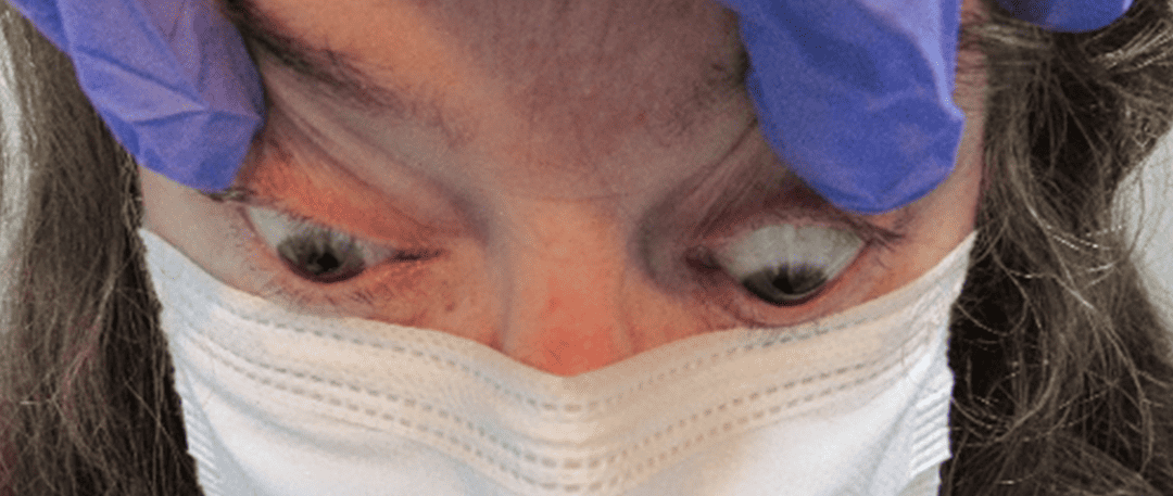
An optometrically driven approach: diagnosing a patient with active thyroid eye disease in the setting of Hashimoto’s thyroiditis
A 57-year-old white female presented with a complaint of intermittent binocular horizontal diplopia with dull ache behind her left eye, described as dull…

Atypical optic neuritis resulting in a diagnosis of MOGAD
The most common systemic cause of optic neuritis continues to be multiple sclerosis.1 However, within the past decade other less common causes have been identified, such as neuromyelitis optica spectrum disorder and myelin oligodendrocyte antibody-associated disease. Previously, patients presenting with these conditions were likely grouped into having multiple sclerosis. Today, thankfully, advancements in laboratory testing and increased knowledge of the central nervous system have allowed them to be identified as distinct nosological entities. Identifying the cause of optic neuritis has many important implications, namely the ability to give an accurate prognosis, both visually and systemically.

Asymptomatic Occipital Stroke Uncovered During Work-up For Optic Neuropathy
Shock-induced ischemic optic neuropathy is often associated with prior gastrointestinal bleeds, and patients with optic disc pallor should be…

Editor’s Column #1
Welcome to the first edition of the Journal of Medical Optometry! JoMO is the official journal of the American Board of Certification in Medical Optometry and we are so proud to bring this publication to you. The genesis of this journal is the attempted solution to a problem: the dearth of journals designed for clinical optometrists.
Neovascular Glaucoma: A Case Report and Literature Review
Neovascular glaucoma is a unique, severe form of glaucoma arising from retinal ischemia. It is often a product of ocular ischemic disorders, such as diabetic retinopathy, ocular ischemic syndrome, and central retinal vein occlusion, all of which may be caused by serious systemic disorders. Neovascular glaucoma must be addressed urgently at onset due to its guarded visual prognosis, the accompanying pain due to the rise in intraocular pressure, and to address the significant underlying systemic etiology.
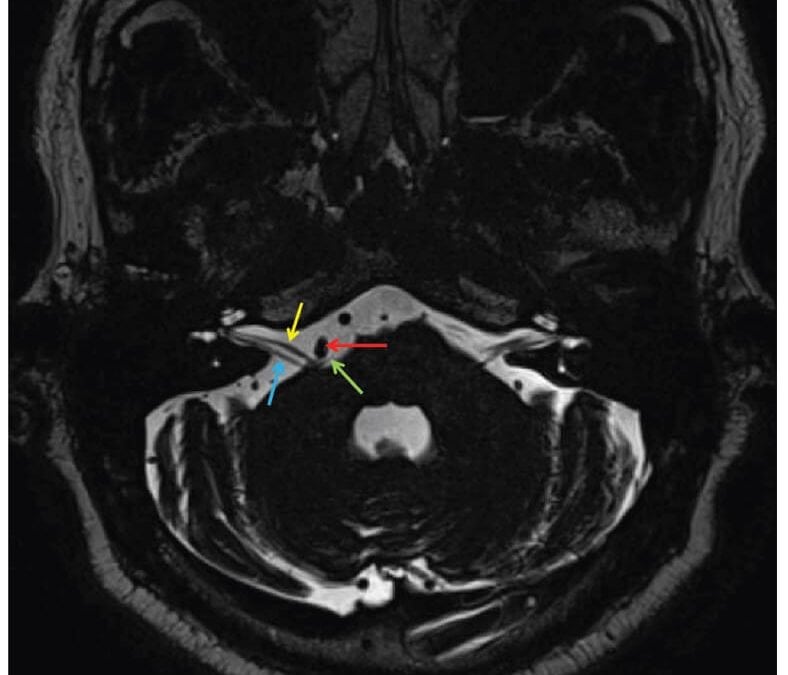
A Multidisciplinary Approach in the Management of Hemifacial Spasm
Patients with hemifacial spasm are often misdiagnosed. This is a case report of a patient with eyelid myokymia who presents with worsening symptoms. Proper diagnosis of hemifacial spasm warrants an MRI revealing: microvascular compression displacing the right 7th cranial nerve at the root exit zone. The role of optometrists and different management options are discussed.
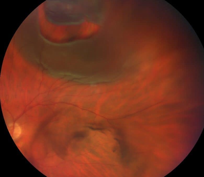
Photo Essay and A Brief Report on New Rhegmatogenous Retinal Detachment Management
This brief report contrasts the differences between pneumatic retinopexy (PnR), pars plana vitrectomy (PPV), and minimal gas vitrectomy (MGV) in rhegmatogenous retinal detachment (RRD) intervention.

Conjunctivochalasis: A Report of Two Cases
Conjunctivochalasis regularly shares symptomatology with multiple other common ocular surface ailments. Additionally, conjunctivochalasis can exist concomitantly with and exacerbate other ocular surface etiologies of discomfort, complicating timely and proper diagnosis and treatment.
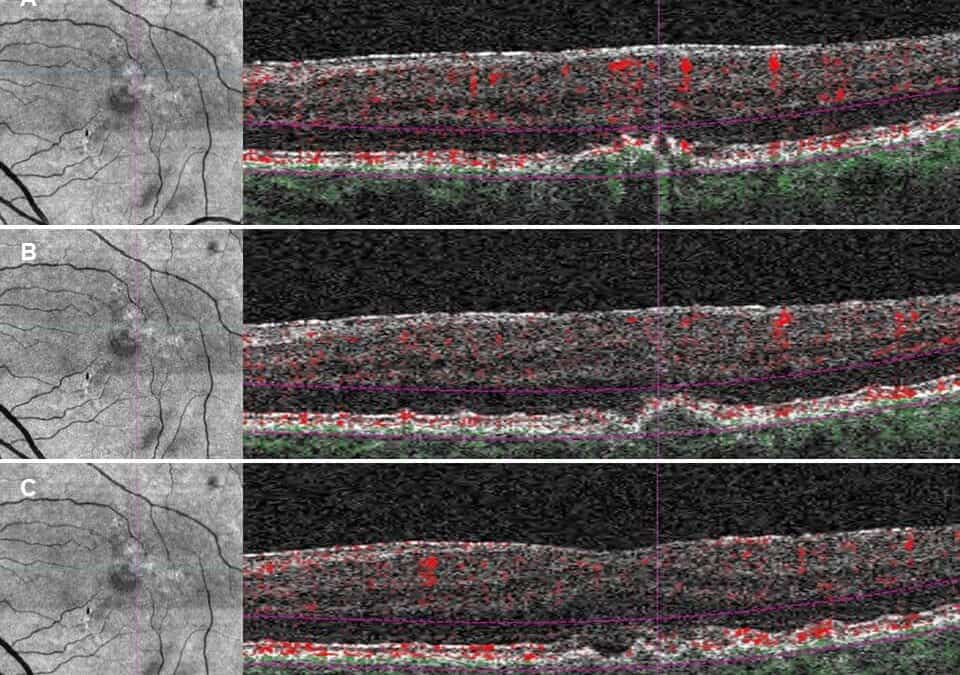
Subjective Dyschromatopsia as presenting symptom of Neovascular Age-Related Macular Degeneration
Subretinal fluid (SRF) is a possible finding associated with neovascular Age-related Macular Degeneration (AMD). SRF has been implicated as a cause of dyschromatopsia in other retinal conditions, such as Central Serous Chorioretinopathy (CSC), but not commonly in AMD. We report a case of sudden onset subjective dyschromatopsia associated with newly noted subretinal fluid secondary to neovascular AMD.

Clinical Decision Making is Difficult
How do we make good decisions for our patients? How do we decide which diagnosis is most appropriate? How do we decide which treatment plan provides the best possible outcome? More fundamentally, how do we decide while uncertain?
An optometrically driven approach: diagnosing a patient with active thyroid eye disease in the setting of Hashimoto’s thyroiditis
A 57-year-old white female presented with a complaint of intermittent binocular horizontal diplopia with dull ache behind her left eye, described as dull…
Atypical optic neuritis resulting in a diagnosis of MOGAD
The most common systemic cause of optic neuritis continues to be multiple sclerosis.1 However, within the past decade other less common causes have been identified, such as neuromyelitis optica spectrum disorder and myelin oligodendrocyte antibody-associated disease. Previously, patients presenting with these conditions were likely grouped into having multiple sclerosis. Today, thankfully, advancements in laboratory testing and increased knowledge of the central nervous system have allowed them to be identified as distinct nosological entities. Identifying the cause of optic neuritis has many important implications, namely the ability to give an accurate prognosis, both visually and systemically.
Asymptomatic Occipital Stroke Uncovered During Work-up For Optic Neuropathy
Shock-induced ischemic optic neuropathy is often associated with prior gastrointestinal bleeds, and patients with optic disc pallor should be…
Editor’s Column #1
Welcome to the first edition of the Journal of Medical Optometry! JoMO is the official journal of the American Board of Certification in Medical Optometry and we are so proud to bring this publication to you. The genesis of this journal is the attempted solution to a problem: the dearth of journals designed for clinical optometrists.
Neovascular Glaucoma: A Case Report and Literature Review
Neovascular glaucoma is a unique, severe form of glaucoma arising from retinal ischemia. It is often a product of ocular ischemic disorders, such as diabetic retinopathy, ocular ischemic syndrome, and central retinal vein occlusion, all of which may be caused by serious systemic disorders. Neovascular glaucoma must be addressed urgently at onset due to its guarded visual prognosis, the accompanying pain due to the rise in intraocular pressure, and to address the significant underlying systemic etiology.
A Multidisciplinary Approach in the Management of Hemifacial Spasm
Patients with hemifacial spasm are often misdiagnosed. This is a case report of a patient with eyelid myokymia who presents with worsening symptoms. Proper diagnosis of hemifacial spasm warrants an MRI revealing: microvascular compression displacing the right 7th cranial nerve at the root exit zone. The role of optometrists and different management options are discussed.
Photo Essay and A Brief Report on New Rhegmatogenous Retinal Detachment Management
This brief report contrasts the differences between pneumatic retinopexy (PnR), pars plana vitrectomy (PPV), and minimal gas vitrectomy (MGV) in rhegmatogenous retinal detachment (RRD) intervention.
Conjunctivochalasis: A Report of Two Cases
Conjunctivochalasis regularly shares symptomatology with multiple other common ocular surface ailments. Additionally, conjunctivochalasis can exist concomitantly with and exacerbate other ocular surface etiologies of discomfort, complicating timely and proper diagnosis and treatment.
Subjective Dyschromatopsia as presenting symptom of Neovascular Age-Related Macular Degeneration
Subretinal fluid (SRF) is a possible finding associated with neovascular Age-related Macular Degeneration (AMD). SRF has been implicated as a cause of dyschromatopsia in other retinal conditions, such as Central Serous Chorioretinopathy (CSC), but not commonly in AMD. We report a case of sudden onset subjective dyschromatopsia associated with newly noted subretinal fluid secondary to neovascular AMD.
Clinical Decision Making is Difficult
How do we make good decisions for our patients? How do we decide which diagnosis is most appropriate? How do we decide which treatment plan provides the best possible outcome? More fundamentally, how do we decide while uncertain?
An optometrically driven approach: diagnosing a patient with active thyroid eye disease in the setting of Hashimoto’s thyroiditis
A 57-year-old white female presented with a complaint of intermittent binocular horizontal diplopia with dull ache behind her left eye, described as dull…
Atypical optic neuritis resulting in a diagnosis of MOGAD
The most common systemic cause of optic neuritis continues to be multiple sclerosis.1 However, within the past decade other less common causes have been identified, such as neuromyelitis optica spectrum disorder and myelin oligodendrocyte antibody-associated disease. Previously, patients presenting with these conditions were likely grouped into having multiple sclerosis. Today, thankfully, advancements in laboratory testing and increased knowledge of the central nervous system have allowed them to be identified as distinct nosological entities. Identifying the cause of optic neuritis has many important implications, namely the ability to give an accurate prognosis, both visually and systemically.
Asymptomatic Occipital Stroke Uncovered During Work-up For Optic Neuropathy
Shock-induced ischemic optic neuropathy is often associated with prior gastrointestinal bleeds, and patients with optic disc pallor should be…
Editor’s Column #1
Welcome to the first edition of the Journal of Medical Optometry! JoMO is the official journal of the American Board of Certification in Medical Optometry and we are so proud to bring this publication to you. The genesis of this journal is the attempted solution to a problem: the dearth of journals designed for clinical optometrists.
Neovascular Glaucoma: A Case Report and Literature Review
Neovascular glaucoma is a unique, severe form of glaucoma arising from retinal ischemia. It is often a product of ocular ischemic disorders, such as diabetic retinopathy, ocular ischemic syndrome, and central retinal vein occlusion, all of which may be caused by serious systemic disorders. Neovascular glaucoma must be addressed urgently at onset due to its guarded visual prognosis, the accompanying pain due to the rise in intraocular pressure, and to address the significant underlying systemic etiology.
A Multidisciplinary Approach in the Management of Hemifacial Spasm
Patients with hemifacial spasm are often misdiagnosed. This is a case report of a patient with eyelid myokymia who presents with worsening symptoms. Proper diagnosis of hemifacial spasm warrants an MRI revealing: microvascular compression displacing the right 7th cranial nerve at the root exit zone. The role of optometrists and different management options are discussed.
Photo Essay and A Brief Report on New Rhegmatogenous Retinal Detachment Management
This brief report contrasts the differences between pneumatic retinopexy (PnR), pars plana vitrectomy (PPV), and minimal gas vitrectomy (MGV) in rhegmatogenous retinal detachment (RRD) intervention.
Conjunctivochalasis: A Report of Two Cases
Conjunctivochalasis regularly shares symptomatology with multiple other common ocular surface ailments. Additionally, conjunctivochalasis can exist concomitantly with and exacerbate other ocular surface etiologies of discomfort, complicating timely and proper diagnosis and treatment.
Subjective Dyschromatopsia as presenting symptom of Neovascular Age-Related Macular Degeneration
Subretinal fluid (SRF) is a possible finding associated with neovascular Age-related Macular Degeneration (AMD). SRF has been implicated as a cause of dyschromatopsia in other retinal conditions, such as Central Serous Chorioretinopathy (CSC), but not commonly in AMD. We report a case of sudden onset subjective dyschromatopsia associated with newly noted subretinal fluid secondary to neovascular AMD.
Clinical Decision Making is Difficult
How do we make good decisions for our patients? How do we decide which diagnosis is most appropriate? How do we decide which treatment plan provides the best possible outcome? More fundamentally, how do we decide while uncertain?
An optometrically driven approach: diagnosing a patient with active thyroid eye disease in the setting of Hashimoto’s thyroiditis
A 57-year-old white female presented with a complaint of intermittent binocular horizontal diplopia with dull ache behind her left eye, described as dull…
Atypical optic neuritis resulting in a diagnosis of MOGAD
The most common systemic cause of optic neuritis continues to be multiple sclerosis.1 However, within the past decade other less common causes have been identified, such as neuromyelitis optica spectrum disorder and myelin oligodendrocyte antibody-associated disease. Previously, patients presenting with these conditions were likely grouped into having multiple sclerosis. Today, thankfully, advancements in laboratory testing and increased knowledge of the central nervous system have allowed them to be identified as distinct nosological entities. Identifying the cause of optic neuritis has many important implications, namely the ability to give an accurate prognosis, both visually and systemically.
Asymptomatic Occipital Stroke Uncovered During Work-up For Optic Neuropathy
Shock-induced ischemic optic neuropathy is often associated with prior gastrointestinal bleeds, and patients with optic disc pallor should be…
Editor’s Column #1
Welcome to the first edition of the Journal of Medical Optometry! JoMO is the official journal of the American Board of Certification in Medical Optometry and we are so proud to bring this publication to you. The genesis of this journal is the attempted solution to a problem: the dearth of journals designed for clinical optometrists.
Neovascular Glaucoma: A Case Report and Literature Review
Neovascular glaucoma is a unique, severe form of glaucoma arising from retinal ischemia. It is often a product of ocular ischemic disorders, such as diabetic retinopathy, ocular ischemic syndrome, and central retinal vein occlusion, all of which may be caused by serious systemic disorders. Neovascular glaucoma must be addressed urgently at onset due to its guarded visual prognosis, the accompanying pain due to the rise in intraocular pressure, and to address the significant underlying systemic etiology.
A Multidisciplinary Approach in the Management of Hemifacial Spasm
Patients with hemifacial spasm are often misdiagnosed. This is a case report of a patient with eyelid myokymia who presents with worsening symptoms. Proper diagnosis of hemifacial spasm warrants an MRI revealing: microvascular compression displacing the right 7th cranial nerve at the root exit zone. The role of optometrists and different management options are discussed.
Photo Essay and A Brief Report on New Rhegmatogenous Retinal Detachment Management
This brief report contrasts the differences between pneumatic retinopexy (PnR), pars plana vitrectomy (PPV), and minimal gas vitrectomy (MGV) in rhegmatogenous retinal detachment (RRD) intervention.
Conjunctivochalasis: A Report of Two Cases
Conjunctivochalasis regularly shares symptomatology with multiple other common ocular surface ailments. Additionally, conjunctivochalasis can exist concomitantly with and exacerbate other ocular surface etiologies of discomfort, complicating timely and proper diagnosis and treatment.
Subjective Dyschromatopsia as presenting symptom of Neovascular Age-Related Macular Degeneration
Subretinal fluid (SRF) is a possible finding associated with neovascular Age-related Macular Degeneration (AMD). SRF has been implicated as a cause of dyschromatopsia in other retinal conditions, such as Central Serous Chorioretinopathy (CSC), but not commonly in AMD. We report a case of sudden onset subjective dyschromatopsia associated with newly noted subretinal fluid secondary to neovascular AMD.
Clinical Decision Making is Difficult
How do we make good decisions for our patients? How do we decide which diagnosis is most appropriate? How do we decide which treatment plan provides the best possible outcome? More fundamentally, how do we decide while uncertain?
An optometrically driven approach: diagnosing a patient with active thyroid eye disease in the setting of Hashimoto’s thyroiditis
A 57-year-old white female presented with a complaint of intermittent binocular horizontal diplopia with dull ache behind her left eye, described as dull…
Atypical optic neuritis resulting in a diagnosis of MOGAD
The most common systemic cause of optic neuritis continues to be multiple sclerosis.1 However, within the past decade other less common causes have been identified, such as neuromyelitis optica spectrum disorder and myelin oligodendrocyte antibody-associated disease. Previously, patients presenting with these conditions were likely grouped into having multiple sclerosis. Today, thankfully, advancements in laboratory testing and increased knowledge of the central nervous system have allowed them to be identified as distinct nosological entities. Identifying the cause of optic neuritis has many important implications, namely the ability to give an accurate prognosis, both visually and systemically.
Asymptomatic Occipital Stroke Uncovered During Work-up For Optic Neuropathy
Shock-induced ischemic optic neuropathy is often associated with prior gastrointestinal bleeds, and patients with optic disc pallor should be…
Editor’s Column #1
Welcome to the first edition of the Journal of Medical Optometry! JoMO is the official journal of the American Board of Certification in Medical Optometry and we are so proud to bring this publication to you. The genesis of this journal is the attempted solution to a problem: the dearth of journals designed for clinical optometrists.
Neovascular Glaucoma: A Case Report and Literature Review
Neovascular glaucoma is a unique, severe form of glaucoma arising from retinal ischemia. It is often a product of ocular ischemic disorders, such as diabetic retinopathy, ocular ischemic syndrome, and central retinal vein occlusion, all of which may be caused by serious systemic disorders. Neovascular glaucoma must be addressed urgently at onset due to its guarded visual prognosis, the accompanying pain due to the rise in intraocular pressure, and to address the significant underlying systemic etiology.
A Multidisciplinary Approach in the Management of Hemifacial Spasm
Patients with hemifacial spasm are often misdiagnosed. This is a case report of a patient with eyelid myokymia who presents with worsening symptoms. Proper diagnosis of hemifacial spasm warrants an MRI revealing: microvascular compression displacing the right 7th cranial nerve at the root exit zone. The role of optometrists and different management options are discussed.
Photo Essay and A Brief Report on New Rhegmatogenous Retinal Detachment Management
This brief report contrasts the differences between pneumatic retinopexy (PnR), pars plana vitrectomy (PPV), and minimal gas vitrectomy (MGV) in rhegmatogenous retinal detachment (RRD) intervention.
Conjunctivochalasis: A Report of Two Cases
Conjunctivochalasis regularly shares symptomatology with multiple other common ocular surface ailments. Additionally, conjunctivochalasis can exist concomitantly with and exacerbate other ocular surface etiologies of discomfort, complicating timely and proper diagnosis and treatment.
Subjective Dyschromatopsia as presenting symptom of Neovascular Age-Related Macular Degeneration
Subretinal fluid (SRF) is a possible finding associated with neovascular Age-related Macular Degeneration (AMD). SRF has been implicated as a cause of dyschromatopsia in other retinal conditions, such as Central Serous Chorioretinopathy (CSC), but not commonly in AMD. We report a case of sudden onset subjective dyschromatopsia associated with newly noted subretinal fluid secondary to neovascular AMD.
Clinical Decision Making is Difficult
How do we make good decisions for our patients? How do we decide which diagnosis is most appropriate? How do we decide which treatment plan provides the best possible outcome? More fundamentally, how do we decide while uncertain?
An optometrically driven approach: diagnosing a patient with active thyroid eye disease in the setting of Hashimoto’s thyroiditis
A 57-year-old white female presented with a complaint of intermittent binocular horizontal diplopia with dull ache behind her left eye, described as dull…
Atypical optic neuritis resulting in a diagnosis of MOGAD
The most common systemic cause of optic neuritis continues to be multiple sclerosis.1 However, within the past decade other less common causes have been identified, such as neuromyelitis optica spectrum disorder and myelin oligodendrocyte antibody-associated disease. Previously, patients presenting with these conditions were likely grouped into having multiple sclerosis. Today, thankfully, advancements in laboratory testing and increased knowledge of the central nervous system have allowed them to be identified as distinct nosological entities. Identifying the cause of optic neuritis has many important implications, namely the ability to give an accurate prognosis, both visually and systemically.
Asymptomatic Occipital Stroke Uncovered During Work-up For Optic Neuropathy
Shock-induced ischemic optic neuropathy is often associated with prior gastrointestinal bleeds, and patients with optic disc pallor should be…
Editor’s Column #1
Welcome to the first edition of the Journal of Medical Optometry! JoMO is the official journal of the American Board of Certification in Medical Optometry and we are so proud to bring this publication to you. The genesis of this journal is the attempted solution to a problem: the dearth of journals designed for clinical optometrists.
Neovascular Glaucoma: A Case Report and Literature Review
Neovascular glaucoma is a unique, severe form of glaucoma arising from retinal ischemia. It is often a product of ocular ischemic disorders, such as diabetic retinopathy, ocular ischemic syndrome, and central retinal vein occlusion, all of which may be caused by serious systemic disorders. Neovascular glaucoma must be addressed urgently at onset due to its guarded visual prognosis, the accompanying pain due to the rise in intraocular pressure, and to address the significant underlying systemic etiology.
A Multidisciplinary Approach in the Management of Hemifacial Spasm
Patients with hemifacial spasm are often misdiagnosed. This is a case report of a patient with eyelid myokymia who presents with worsening symptoms. Proper diagnosis of hemifacial spasm warrants an MRI revealing: microvascular compression displacing the right 7th cranial nerve at the root exit zone. The role of optometrists and different management options are discussed.
Photo Essay and A Brief Report on New Rhegmatogenous Retinal Detachment Management
This brief report contrasts the differences between pneumatic retinopexy (PnR), pars plana vitrectomy (PPV), and minimal gas vitrectomy (MGV) in rhegmatogenous retinal detachment (RRD) intervention.
Conjunctivochalasis: A Report of Two Cases
Conjunctivochalasis regularly shares symptomatology with multiple other common ocular surface ailments. Additionally, conjunctivochalasis can exist concomitantly with and exacerbate other ocular surface etiologies of discomfort, complicating timely and proper diagnosis and treatment.
Subjective Dyschromatopsia as presenting symptom of Neovascular Age-Related Macular Degeneration
Subretinal fluid (SRF) is a possible finding associated with neovascular Age-related Macular Degeneration (AMD). SRF has been implicated as a cause of dyschromatopsia in other retinal conditions, such as Central Serous Chorioretinopathy (CSC), but not commonly in AMD. We report a case of sudden onset subjective dyschromatopsia associated with newly noted subretinal fluid secondary to neovascular AMD.
Clinical Decision Making is Difficult
How do we make good decisions for our patients? How do we decide which diagnosis is most appropriate? How do we decide which treatment plan provides the best possible outcome? More fundamentally, how do we decide while uncertain?